Prior to working on living beings or attempting more complex procedures as in surgery, the student needs to develop fundamental dexterity or psychomotor skills. Inanimate objects provide a non-stressful, time-efficient and highly effective means of obtaining these skills. Some of these can also be considered as a means of surgical instruction and may be listed in that section. Following the list of specific alternatives, there is a section on literature that either explains or evaluates the alternatives or provides additional information on the subject of skills development.
If you are aware of other examples you believe to be important to include here, please send the information to HEVM for consideration.
New additions:
Validation Studies Require Student Scores and Are Not Necessary for Every Clinical Skills Model
Development and Validation of a Bovine Coccygeal Venipuncture Model and Rubric
 |
 |
 |
Available from Limbs & Things.
From Web site: A comprehensive trainer for teaching all surgical knot tying techniques…
Suture training pad
 |
Available through Veterinary Simulator Industries Ltd.
Skin Suture Pattern Simulator
 |
See more information on the Norecopa site.
Skin Closure Simulator
 |
Produced by Delletec Surgical Procedure Simulators. As of 2023-12-10, however, the link provided for the product is no longer valid. You would need to contact the company directly to find out more information.
 |
 |
 |
Produced by Rescue Critters!.
Adjustable Tissue Tray Package
 |
 |
For suturing skin incisions. The incision can be adjusted to create different degrees of tension.
Produced by Simulab Corporation.
 |
 |
 |
Available through Paws 2 Claws™.
From Web site: A great tool to learn or practice sutures on. One side includes a representation of a canine elbow bone.
 |
Produced by Simulab Corporation.
 |
 |
 |
For practicing blood vessel ligation.
Produced by Rescue Critters!.
RealLayer RealFlow simulated tissue technology
 |
 |
 |
For hemostasis and suturing. The company produces several simulators for practicing suturing.
Produced by SurgiReal.
Hemostasis model
 |
 |
 |
By Olsen et al 1996: Results of this study indicate that the hemostasis model was as effective as live animals for teaching the basic skills involved in blood vessel ligation. The students’ opinions regarding the use of properly designed inanimate models for teaching these skills were dramatically changed.
Foam hemostasis model
 |
 |
 |
By Smeak et al 1991: Students using simulators performed ligation with significantly more accuracy and tended to be more expeditious at this task.
Hemostasis simulator
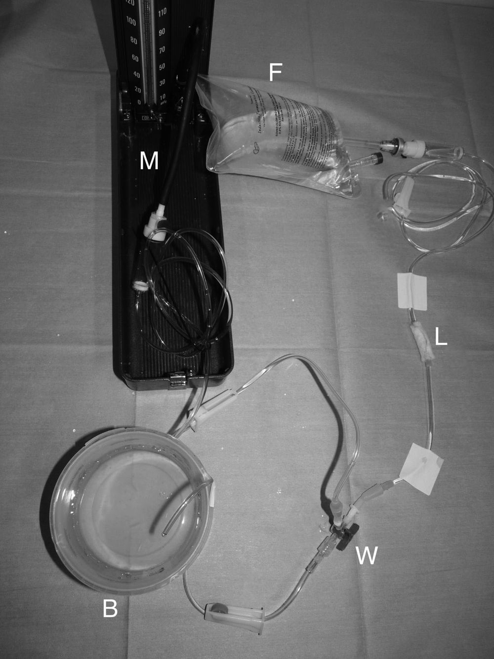 |
See Giusto et al 2015 for details and results of assessment.
 |
 |
 |
Produced by Simulab Corporation.
From Web site: The laparotomy model simulates a partial abdomen and includes skin, subcutaneous fat, fascia, preperitoneal fat, and peritoneum.
Hollow Organ Simulator
 |
Reviewed by Smeak et al 1994.
 |
Produced by SurgiReal.
This is a small intestine simulator for the practice of end to end, side to side, end to side and functional end to end anastomotic techniques.
Double Layer Bowel 20mm Outside Diameter
 |
Available from Limbs & Things.
From Web site: Realistic two layer bowel simulation for training in anastomosis techniques. … Realistic tissue response … Will withstand a fluid flush test to demonstrate integrity of the anastomosis
 |
Produced by Simulab Corporation.
From Web site: The Double-layered Intestine is a bowel section with two distinct layers for increased realism. Use this product to practice suturing, anastomosis, and stapling.
Vascular Access Training Models
Canine foreleg model |
Canine head and foreleg model |
Used for training in the art of vascular access (phlebotomy).
They were originally produced and sold by the University of California School of Veterinary Medicine, but the original page is no longer available. You can contact the university directly to see if there is more information.
Canine Leg Vascular Access Simulator
 |
 |
 |
 |
Vascular access simulator for the dog.
Produced by SurgiReal.
Horse jugular vein access model
 |
Described and evaluated in Eichel et al 2013.
 |
 |
Vascular access simulator for the horse; jugular and facial arteries.
Produced by SurgiReal.
Equine phlebotomy and intramuscular injection simulator
 |
Vascular and muscle access for the horse.
This simulator is described in by Williamson et al 2016.
Intravenous cannulation simulator
 |
Students using this easily made simulator showed more skill in cannulating the vein of a live animal than those not practicing on the simulator first, as reported by Perez-Rivero & Rendón-Franco 2011.
Alpaca vascular access
 |
This model was evaluated by Rousseau et al 2017.
Dog cadaver leg for venipuncture
 |
Galle & Bubna-Littitz 1983 used a formalin-fixed front leg of a dog and replaced the cephalic vein with a silicon tube containing artificial blood. This was then used to teach students the art of venipuncture. With ethically-sourced cadavers, this could be an effective means of training.
Fine needle aspiration device
 |
Developed by Simpson & Meuten 1992. This simple device, described in the publication, is used to contain fresh organ material (obtained from the necropsy lab) for teaching students the art of fine needle aspiration.
DASIE™ Dog Abdominal Surrogate for Instructional Exercise
 |
See Holmberg et al 1993 for evaluation.
Dog abdomen, ovariohysterectomy simulator
 |
 |
 |
This appears to be an effective means of preparing the student for ovariohysterectomy in the living animal.
Developed by University of Sydney, Faculty of Veterinary Science, Sydney, Australia with Studio Kite; see article by Woon 2011 for more information.
TheMOOSE, ovariohysterectomy simulator
 |
This is described and evaluated by Fahie et al 2016.
Spay Training Manikin (ovariohysterectomy)
 |
 |
Produced by Vet Effects Incorporated.
From Web site: The manikin allows the student to recreate a lifelike, step by step training, from incision to sutures.
Manikin – Spay Dog (ovariohysterectomy)
 |
 |
 |
 |
Available through Paws 2 Claws™.
From Web site: The Spay Manikin was designed by Paws 2 Claws to be hands on training aid in learning the surgical procedures and techniques of spaying a dog, from intubation to the final sutures.
Neuter Training Manikin (castration)
 |
 |
Produced by Vet Effects Incorporated.
From Web site: The manikin allows the student to recreate a lifelike, step by step training, from incision to sutures.
Manikin – Neuter Dog (castration)
 |
 |
 |
 |
 |
Available through Paws 2 Claws™.
From Web site: The Neuter Manikin was designed by Paws 2 Claws to be hands on training aid in learning the surgical procedures and techniques of neutering a dog, from intubation to the final sutures.
Various simulators at Utrecht University
Cat castration |
Dog castration |
Tracheostomy |
Venipuncture |
Drs van Nimwegen and Kirpensteijn at Utrecht University have developed a psychomotor skills program which includes various skills models or simulators. Although the simulators may appear ‘primitive’, they should be useful in developing initial dexterity for a wide range of procedures including cat castration, pre-scrotal castration of the dog, tracheostomy tube placement, pedicle ligation in ovariohysterectomy, esophageal feeding tube placement, venipuncture, tumor excision.
You will need to contact the university or the authors to find out if there is any more information about what the authors have developed: Utrecht University, Faculty of Veterinary Medicine
 |
Produced by Rescue Critters!.
From Web site: …performing chest tube placements, and simulating emergency trauma by aspirating air and fluid from the thoracic cavity.
Training model for small animal thoracocentesis and chest tube thoracostomy
 |
By Williamson & Fio Rito 2014: A small animal thoracocentesis and chest tube thoracostomy model has been developed, that allows repetitive practice in a safe, standardised environment. … Student feedback indicated a high degree of satisfaction with the model and the laboratory experience, high perceived value of the case studies in improving learning, and increased confidence to perform the procedures under supervision. This model can replace the use of live animals while students are practising these procedures, improving their technique, and learning the appropriate safeguards used to prevent injuries such as pulmonary trauma.
See Williamson 2014 for critical evaluation.
 |
 |
 |
Produced by Rescue Critters!.
 |
 |
Produced by Rescue Critters!
From Web site: Representations of the trachea, esophagus and epiglottis are all realistically crafted into this dog bust. It also features an airway with working lungs.
 |
Produced by Nasco.
 |
Produced by Rescue Critters!.
From Web site: Like its canine counterpart CCJ, Critical Care Fluffy is a full-sized, realistic feline mannikin.
 |
Produced by Rescue Critters!.
From Web site: …engineered as a complete emergency room veterinary training mannikin.
Low-fidelity canine models for intubation and urinary catheterization
 |
 |
Described and evaluated in Aulmann et al 2015.
Human patient simulator
 |
Modell et al 2014 used a human patient simulator from CAE Healthcare to train veterinary medical students in anesthesia and complications that may occur. They concluded that …the human patient simulator was a valuable learning tool for students of veterinary medicine. It was exciting for the students to work with, made them deal with ‘real-life’ scenarios, permitted them to learn without subjecting live patients to complications, enabled them to retrace their steps when their therapy did not correct the simulated patient’s problems, and facilitated correlation of their basic science knowledge with clinical data, thus accelerating their ability to handle complex clinical problems in healthy and diseased patients.
 |
 |
Developed by Dr Daniel Fletcher at Cornell University College of Veterinary Medicine. There also is a similar simulator for cats. All the simulators are stated to be available for sharing with other institutions. The dog simulator is described further by Fletcher et al 2012.
Flexible and Rigid Endoscopic training Device (FRED)
") |
") |
Developed by Jacqueline C Whittemore, DVM, PhD, DACVIM at the University of Tennessee College of Veterinary Medicine. See Kennedy 2009 for news item.
Simuldog
 |
 |
 |
Development, use and validation reported in Usón-Gargallo et al 2014.
Additional evaluation by Pérez-Merino et al 2018.
Canine Laparoscopic Ovariectomy Model
Described by Chen et al 2019.
Canine Laparoscopic Simulator
 |
 |
 |
Development, use and validation reported in Usón-Gargallo et al 2014.
 |
Available from SynDaver Labs.
From Web site: The SynDaver Synthetic Canine is a futuristic animal model designed to replace live animals and animal cadavers in veterinary surgical training. Based on 20+ years of SynDaver research, this model is made from water, fiber and salt. She is a life saver, but she is not alive. She breathes and bleeds just like a real dog. She has individual muscles, bones, and organs – and can be operated on repeatedly without risking a live animal.
Joint Injection Simulator
 |
Described and evaluated in Fox et al 2013.
Cadaver model for arthrocentesis
Described and evaluated in MacIver & Johnson 2015.
Canine pelvic limb model for stifle joint evaluation
 |
 |
Described and evaluated in Troy & Bergh 2015.
Bovine rectal palpation simulator
 |
 |
Uses haptic technology. Developed at the University of Glasgow Veterinary School, as reported by Baillie et al 2005 and validated in Baillie et al 2005, and Kinnison et al 2009. An automated version was developed and evaluated by Baillie et al 2010.
 |
Report on its use and instructional evaluation in Read & Baillie 2013.
 |
Its use and evaluation were reported in Bossaert et al 2009.
Equine Palpation Colic Simulator
 |
For practicing rectal palpation in the female horse.
Available through Veterinary Simulator Industries Ltd.
Horse Ovary Palpation Simulator
 |
As reported by Crossan et al.
Canine prostate palpation simulator
 |
Described and evaluated in Capilé et al 2015.
Male Urinary Catheter Training Manikin
 |
Available through Paws 2 Claws™.
From Web site: This manikin was created to simulate the urinary catheterization procedure, along with collecting urine, emptying the bladder or taking urine samples.
 |
 |
 |
 |
Available through Paws 2 Claws™.
From Web site: This manikin is designed with an external and internal urogenital structure with the purpose of performing two separate procedures; cystocentesis and cystostomy.
There is also the Cysto Dog Manikin.
Dental model for training veterinary and veterinary nursing students
 |
 |
 |
This is a being used at the Royal Veterinary College …to facilitate the teaching, learning and assessment of basic dental skills for veterinary nursing and veterinary medicine students.
An evaluation of it was reported by Lumbis et al 2012.
Rabbit silicon ear
 |
 |
 |
For intravascular access practice in the rabbit.
Produced by NPM SIKO.
 |
 |
 |
For practicing endotracheal intubation, cardiac puncture, blood collection from the saphenous vein in the rat.
Produced by Rescue Critters!.
 |
From Web site: …silicone rat designed for use on medical, pharmaceutical and veterinary training courses.
Koken Rat
 |
For practicing restraint, peroral dosing, intravenous vascular access through tail, endotracheal intubation in the rat.
There is purported to be a Koken Rabbit which is used for practicing restraint, peroral dosing, vascular access using the auricular vein, endotracheal intubation, urine collection through urethral catheterization.
Canine eye model for ophthalmoscopy
 |
Although ophthalmoscopy is not particularly invasive and student or staff companion animals can be used without any harm to them, this model may at least allow students to develop the psychomotor skills necessary without the added struggle of a moving “patient”.
Described and evaluated in Nibblett et al 2015.
Bovine Birthing and Ultrasound Simulator

Bovine Injection Simulator

Calf Simulator

The following includes literature cited above or which is relevant to the development of various skills, including outcome assessment. The titles are linked either to a publicly available copy of the document or to a digital object identifier. If there are illustrations which may be publicly viewable, these are also linked, but there is no guarantee that they would be viewable across all platforms.
Abutarbush, Sameeh M. et al
2006
Evaluation of traditional instruction versus a self-learning computer module in teaching veterinary students how to pass a nasogastric tube in the horse
Journal of Veterinary Medical Education 33(3):447-454
Conclusion – computer-assisted learning is an acceptable and effective method of training students to pass an NG tube with potential welfare, proficiency, and knowledge advantages.
Article is stated to be open access. Alternately, you may be able to obtain a copy of the paper from: ResearchGate
Allen, S.W. et al
1997
Computer-assisted instruction of fundamental surgical motor skills
Journal of Veterinary Medical Education 24(1):2-5
A computer-assisted learning program, ‘The Surgical Techniques Auto-Tutorial Program,’ was developed for use as an introductory training tool of fundamental surgical motor skills. The program was well received by veterinary medical students. Although computer-assisted instruction was as effective as traditional methods in helping students develop and retain some skill, direct instructor contact was necessary for the retention of other stills such as knot tying. When followed by instructor contact laboratories, allowing feedback and reinforcement of operative skills, computer-assisted instruction was a helpful introductory training tool for the development of fundamental surgical motor skills.
Anderson, Lane S. et al
2023-12-01
Proficiency and Retention of Five Clinical Veterinary Skills Using Multipurpose Reusable Canine Manikins versus Live Animals: Model Development and Validation
Journal of Veterinary Medical Education 50(6):654-660
 |
 |
 |
 |
 |
…canine training manikins were created using readily available materials to teach fine needle aspiration (FNA) of peripheral lymph nodes, jugular venipuncture, cephalic venipuncture, intravenous catheterization, and cystocentesis…Initial proficiency and short-term retention of clinical skills do not differ for students trained using a manikin versus a live dog.
Article is stated to be open access.
Annandale, Annett et al
2018-06-01
Training method and other factors affecting student accuracy in bovine pregnancy diagnosis
Journal of Veterinary Medical Education 45(2):224-231.
Annandale, Annett et al
2020
The effect of an ovariohysterectomy model practice on surgical times for final-year veterinary students’ first live-animal ovariohysterectomies
Journal of Veterinary Medical Education 47(1):44-55
 |
 |
…students practicing an OVH on the model felt more confident (92%) and less stressed (73%) when performing their first live-animal OVH. Results suggest that the canine OVH model may be helpful as a clinical training tool and we concluded that the OVH model was effective at decreasing students’ first OVH surgical time.
Article is stated to be open access.
Anonymous
2024-10-10
3D-printed model helps train neonatal kitten feeding
Vet Times
 |
Led by Karen Vernau and individuals from the Translating Engineering Advances to Medicine (TEAM) Lab at the university, the project has resulted in lifelike, 3D-printed kitten models being designed to improve hands-on training for veterinary students and caregivers.
The team produced a silicone model that includes a trachea and oesophagus, designed to provide immediate feedback if a tube is inserted incorrectly, with a unique Y-shaped fork mechanism that prompts users to correct their technique.
It has an embedded 3D-printed ribcage, which serves as a crucial anatomical landmark to help users gauge the correct length of the feeding tube.
Auer, Jörg Andreas
1994
Veterinär-Chirurgische Ausbildung am Simulator
ALTEX 11(1):44-46
Discusses the use of simulators for development of basic surgical skills at the Veterinary Surgery Clinic of the University of Zürich. In German with an English summary.
Aulmann, Maria et al
2015
Development and evaluation of two canine low-fidelity simulation models
Journal of Veterinary Medical Education 42(2):151-160
 |
 |
 |
We thereby conclude that low-fidelity models can be as effective as high-fidelity models for clinical skills training.
Article is stated to be open access. Alternately, you may be able to obtain a copy of the paper from: ResearchGate
Baillie, S.
Utilization of simulators in veterinary training
15 pp
Baillie, Sarah et al
2005Integrating a bovine rectal palpation simulator into an undergraduate veterinary curriculum
Journal of Veterinary Medical Education 32(1):79-85
Baillie, Sarah et al
2005
Validation of a bovine rectal palpation simulator for training veterinary students
Studies in Health Technology and Informatics 111:33-36
The subsequent performance in the real task, when examining cows for the first time, was assessed with the results showing a significantly better performance for the simulator group.
Baillie, Sarah et al
2010
Evaluating an automated haptic simulator designed for veterinary students to learn bovine rectal palpation
Simulation in Healthcare 5(5):261-266
The automated simulator equipped students with useful skills for examining cows. In addition, a simulator that does not need the presence of an instructor will increase the availability of training for students and be a more sustainable option for institutions.
Baillie, Sarah et al
2020-07-01
Comparison of a Silicon Skin Pad and a Tea Towel as Models for Learning a Simple Interrupted Suture
Journal of Veterinary Medical Education 47(4):516-522
 |
In conclusion, the tea towel was as effective as the silicon skin pad, but it was cheaper, simpler to make, and the materials were more readily available. In addition, both models were used effectively with an instruction booklet illustrating the value of self-directed learning to complement taught classes.
Article is stated to be open access.
Baillie, Sarah et al
2022-12-01
Designing Flipped Classrooms to Enhance Learning in the Clinical Skills Laboratory
Journal of Veterinary Medical Education 49(6):699-704
A well-designed flipped classroom motivates learners by including different elements such as quality educational media (eg, videos), the opportunity to self-assess, and well-defined connections to relevant knowledge and skills. Student engagement with the flipped material can be promoted through different strategies such as clear communication to manage student expectations and adapting the delivery of the face-to-face component. Finally, gathering feedback and evaluating the initiative are important to inform future improvements.
Baillie, Sarah et al
2024-04-01
The Rapid and International Expansion of Veterinary Clinical Skills Laboratories: A Survey to Establish Recent Developments
Journal of Veterinary Medical Education 51(2):215-228
In summary, veterinary clinical skills laboratories are increasingly common around the world and the contributions to student learning and animal welfare were well recognized. The information about existing and planned laboratories and the tips from those managing the facilities provides valuable guidance for anyone intending to open or expand an existing clinical skills laboratory.
Bakici, Caner et al
2026-06-01
Impact of Clinical Skills Laboratory Training and Online Education on Suture Skill Development in Veterinary Students: A Gender-Based Analysis
Journal of Veterinary Medical Education 53(3):396-406
Students who participated in hands-on practice achieved significantly higher post-test scores compared with those who relied solely on online instruction, reinforcing the effectiveness of practical training. Notably, female students in both groups exhibited a statistically higher increase in performance scores than their male counterparts. These findings underscore the importance of practical, model-based training in CSL [clinical skills laboratory] for fostering skills acquisition and revealed the impact of gender on skill development. This study contributes to the growing body of evidence supporting the integration of experiential learning into veterinary education and offers insights into optimizing training methods to enhance student outcomes.
Ball, Leslie et al
1964
An artificial uterus for laboratory instruction
Journal of the American Veterinary Medical Association 144(3):264-265
Describes a model which can be built to teach palpation in the cow.
Banse, Heidi E. et al
2021-10-01
Development of and Validity Evidence for a Canine Ocular Model for Training Novice Veterinary Students to Perform a Fundic Examination
Journal of Veterinary Medical Education 48(5):620-628
 |
Although ophthalmoscopy is not particularly invasive and student or staff companion animals can be used without any harm to them, this model may at least allow students to develop the psychomotor skills necessary without the added struggle of a moving patient.
These findings suggest that this canine model may be an effective tool to train students to perform fundoscopy.
Article is stated to be open access.
Bartner, Howard et al
1971-05-01
An Improved Model for Instruction in Binocular Indirect Ophthalmoscopy
Archives of Ophthalmology 85(5):530-533
Describes a model which can be used to develop skills necessary for this procedure. Although it is for human ophthalmology residency training, could be used in veterinary ophthalmology residency or student training.
Bauer, Michael S.
1992-12-01
Letter to the editor
Journal of Veterinary Medical Education 19(1):32
We believe the creation of lesions suited to the student’s level of training in a setting where preoperative radiographs and discussion are possible, creates a stimulating problem-solving situation that cannot be achieved by use of live animals.
Beaulieu, Alexandra et al
2022-08-01
Development and Validation of a Three-Dimensional Printed Training Model to Teach Ultrasound-Guided Injections of the Cervical Articular Process Joints in Horses
Journal of Veterinary Medical Education 49(4):442-453
 |
 |
 |
 |
…we successfully developed a 3D printed model of an equine cervical articular process joint, partially demonstrated the construct validity of the model, and proved the face and content validity of this new training tool.
Article is stated to be open access.
Bishop, Rebecca C. et al
2025-03-01
How to perform a transtracheal aspirate in horses for diagnosis of lower respiratory tract disease
Journal of the American Veterinary Medical Association 263(3):1
Transtracheal aspirate is minimally invasive and simple to perform with available kits. Samples provide valuable information to guide treatment decisions and selection of antimicrobials for horses with suspected lower respiratory tract disease.
Boger, Brooke L. et al
2024-08-01
Accuracy and Confidence in Performing Canine Stifle Goniometry was Similar between Simulation-Model or Traditional Textbook Trained Veterinary Students
Journal of Veterinary Medical Education 51(4):512-521
 |
 |
 |
In general, learning with models was preferred by all. There was no difference in learning between the model and textbook, so either can be used based on student preference. Further goniometer instructions should be provided. Anatomy of live dogs should be assessed more frequently pre-clinically.
Article is stated to be open access.
Bogert, K. et al
2016
Development and use of an interactive computerized dog model to evaluate cranial nerve knowledge in veterinary students
Journal of Veterinary Medical Education 43(1):26-32
…we have developed a computerized simulated dog head that can exhibit cranial nerve dysfunctions and respond to specific testing procedures in a clinically accurate manner. … In an experiment conducted with 97 freshman veterinary students who had recently been taught cranial nerve anatomy and function, we found that examination performance decreased with the need for interactivity compared to memorization of fact, while satisfaction increased. Students were less likely to identify the correct disorder when they had to conduct the examination of the virtual dog themselves, revealing an inadequacy in traditional neuroanatomical teaching. However, students overwhelmingly supported the use of interactive question for assessment. Interestingly, performance on text-based questions did not correlate significantly with interactive or video questions. The results have implications for veterinary teaching and assessment within the classroom and in clinical environments.
Bonnema, Hannah et al
2026-02-01
Can a Simple Model Have Value Without Validation? A Study to Develop and (Attempt to) Validate a Bovine Caudal Epidural Model and Rubric
Journal of Veterinary Medical Education 53(1):115-122
Bovine practitioners expect new graduates entering clinical practice to be able to place a caudal epidural. Teaching this task on models facilitates scheduled training sessions and sufficient practice to reach competency. This study sought to create and validate a bovine caudal epidural model and scoring rubric…Veterinarians reported that the model was helpful for students to learn and practice the task and that the model had sufficient landmark features and realism…Rubric scores achieved acceptable internal consistency after one item was dropped… and there was no significant difference between veterinarians’ and students’ performance scores on the model…Educators must consider whether simple models that are helpful for students to practice their skills may still have value, even if they are not able to be validated.
Bossaert, Philippe et al
2009
Teaching transrectal palpation of the internal genital organs in cattle
Journal of Veterinary Medical Education 36(4):451-460
 |
Results suggest that Breed’n Betsy cannot fully replace training in live cows, but may be a valuable addition to the classical teaching method. Suggestions for future improvement are made.
Article is stated to be open access.
The Breed’n Betsy Web site is http://www.breednbetsy.com.au/.
Braid, Helen R.
2022-05-01
The Use of Simulators for Teaching Practical Clinical Skills to Veterinary Students — A Review
Alternatives to Laboratory Animals 50(3):184-194
The reviewed articles revealed that there are a number of simulators currently available to veterinary educators, that simulators can enhance student skills and provide an alternative learning environment without the need for live animal and/or cadaver use, and that they usually receive positive feedback from the students who use them. There appears to be a bias towards small animal simulators — however, some skills that are developed through the use of small animal or table-top models will be transferrable to other species. The majority of large animal simulators focus on bovine rectal palpation and/or pregnancy diagnosis.
Brisson, Brigitte A. et al
2023-06-01
Excellent Agreement of In-Person Scoring versus Scoring of Digitally Recorded Simulated Suture Skills Examination
Journal of Veterinary Medical Education 50(3):379-383
 |
The excellent agreement between in-person and digital assessment suggests that digitally recording skills examinations can provide adequate suturing skills assessment, presenting several benefits. Digitally recorded assessment allows for anonymity, which can reduce assessor bias/favoritism, provide a record of performance that students can review and critically self-reflect upon, and reduce the number of in-person examiners required to complete surgical skills examinations. Additionally, digitally recorded assessment could become an option for students who experience anxiety performing a skills exam in the presence of an examiner.
Article is stated to be open access.
Burns, Jeffrey P. et al
1994
Using newly deceased patients to teach resuscitation procedures
The New England Journal of Medicine 331(24):1652-1655
The authors discuss the ethical and legal aspects of this situation, and offer criteria for keeping it a legitimate form of training for the benefit of human society. There is no reason why similar principles could not be applied in veterinary medicine with respect to using new deceased patients for training purposes.
A total of 449 questionnaires were mailed in the summer of 1992, and responses were received from 353 training programs (79 percent). Of these, 136 (39 percent) described using newly deceased patients in the teaching of resuscitation procedures; this finding is similar to the results of another recent survey… The highest proportions of respondents who allowed procedures to be performed on patients after their death were found among the emergency-medicine programs (63 percent) and the neonatal critical care programs (58 percent). Forty percent of programs that used this teaching technique reported using it 10 or more times per year. Tracheal intubation was overwhelmingly the most common procedure practiced, but other procedures, including the placement of central venous catheters, surgical venous cutdown, thoracotomy, pericardiocentesis, cricothyrotomy, liver biopsy, and intraosseous needle placement, were also reported.
Capilé, Karynn V. et al
2015
Canine prostate palpation simulator as a teaching tool in veterinary education
Journal of Veterinary Medical Education 42(2):146-150
 |
…our aim was to develop a canine prostate palpation simulator to provide students with the opportunity to learn the prostate palpation technique in dogs and to assess their opinion of this simulator as a teaching tool. The inner part of the canine mannequin contains a rotation system with three types of prostates that can be exchanged during the exam. … We conclude that the simulator can help students to develop clinical skills for prostate palpation in dogs.
Article is stated to be open access. Alternately, you may be able to obtain a copy of the paper from: ResearchGate
Carroll, Hillary S. et al
2016
Development of an optional clinical skills laboratory for surgical skills training of veterinary students
Journal of the American Veterinary Medical Association 248(6):624-628
During each OCSL session, a variety of surgical training models and cadavers were available for students to practice with. Simple models for students to practice suturing, pedicle ligation, and IV catheter placement were made from surplus hospital stock and inexpensive materials readily available from hardware and craft stores. In addition, 5 to 7 whole or prosected cadavers were typically available for student use during each session. Large animal cadavers were primarily obtained through donation to the veterinary teaching hospital. Canine and feline cadavers were obtained through the CVM’s contract with animal control agencies in Washington state under which animals judged to be unadoptable by individual animal control agencies were euthanized at the agencies in accordance with AVMA and institutional animal care and use guidelines and transported to WSU CVM fresh-frozen. Fresh cadavers from other teaching laboratories were also used when available. All cadaver use adhered to AVMA and institutional animal care and use guidelines for humane use of animals. No animals were euthanized specifically for use in the OCSL.
Chen, Chi-Ya et al
2019
Development and evaluation of a high-fidelity canine laparoscopic ovariectomy model for surgical simulation training and testing
Journal of the American Veterinary Medical Association 254(1):113-123
 |
 |
 |
Results suggested the [simulated laparoscopic ovariectomy] model may be a useful surgical training tool. Further studies are needed to confirm usefulness of the model in veterinary laparoscopy training.
Cosford, Kevin et al
2019
Evaluation of a first-year veterinary surgical skills laboratory: A retrospective review
Journal of Veterinary Medical Education 46(4):423-428
Cosford, Kevin et al
2020-04-01 Effect of instructional format on veterinary students’ task performance and emotional state during a simulation-based canine endotracheal intubation laboratory: Handout versus video
Journal of Veterinary Medical Education 47(2):239-247
Video instructions may be associated with higher performance scores than handout instructions during endotracheal intubation simulation training.
Crossan, Andrew et al
Comparison of simulated ovary palpation training over different skill levels
5 pp University of Glasgow
This paper describes an initial attempt to compare performance levels of users of different skill levels on the Glasgow Horse Ovary Palpation Simulator (HOPS). … Traditionally, students are taught horse ovary palpation through books, lectures, and practical experience. … As ovary palpation is a stressful procedure for the horse, ethical considerations limit a student’s opportunity to gain experience. A horse ovary examination can be difficult for a veterinary student to perform, but can also be fatal to the horse if performed incorrectly.
da Costa, Bruna N. et al
2022-05-01
The Use of 3-D Models of Echocardiographic Imaging Planes for Teaching Echocardiography Techniques for Use in Dogs and Cats
Alternatives to Laboratory Animals 50(3):208-220
The 3-D models facilitated, and significantly improved, the identification of cardiac structures and disease-associated abnormalities, and the learning process in general. Additionally, the models seemed to provide greater student motivation for studying echocardiography.
Dankelman, Jenny
2008
Surgical simulator design and development
World Journal of Surgery 32(2):149-155.
 |
 |
 |
de Solis, Cristobal Navas et al
2021-01-15
Effectiveness of a digital interactive multimedia tutorial for preparing veterinary students to perform ultrasonography in horses
Journal of the American Veterinary Medical Association 258(2):165-169
Higher-quality ultrasound images were obtained by veterinary students who had reviewed the DIMT [digital interactive multimedia tutorial] rather than the analogous information in textbook chapters. No difference in scores was identified between students in the lecture group and those in the DIMT group.
Decloedt, Annelies et al
2021-06-01
Development of Surgical Competence in Veterinary Students Using a Flipped Classroom Approach
Journal of Veterinary Medical Education 48(3):281-288
Flipped classroom CSL [clinical skills laboratory] training resulted in significantly higher self-efficacy (score/100, pre-test 55 ± 14 vs. post-test 83 ± 8, p< .001) and surgical skills performance (score/20, pre-test 5 ± 3 vs. post-test 17 ± 3, p< .001). In conclusion, this study demonstrated the feasibility and value of implementing a flipped classroom approach in combination with CSL training.
Dilly, Marc et al
2017
A survey of established veterinary clinical skills laboratories from Europe and North America: Present practices and recent developments
Journal of Veterinary Medical Education 44(4):580-589
The findings indicated that having a dedicated veterinary clinical skills laboratory is a relatively new initiative and that colleges have adopted a range of approaches to implementing and running the laboratory, teaching, and assessments. Major strengths were the motivation and positive characteristics of the staff involved, providing open access and supporting self-directed learning. … There is no doubt that veterinary clinical skills laboratories are on the increase and provide opportunities to enhance student learning, complement traditional training, and benefit animal welfare.
Dronfield, Amy F. et al
2023-12-01
Comparing the Efficacy of a New Clinical Skills Model with a Traditional Method to Teach Tube Feeding of an Avian Patient
Journal of Veterinary Medical Education 50(6):732-742
…a new model of a bird, made from a soft toy, silicone, and 3D printed parts, was designed to train students to perform this technique…the newly developed model in combination with an instruction booklet offers an effective and inexpensive alternative way to teach crop tubing in a teaching environment, without compromising animal welfare.
Eichel, Jane-Carolin et al
2013
Evaluation of a training model to teach veterinary students a technique for injecting the jugular vein in horses
Journal of Veterinary Medical Education 40(3):288-295
 |
The training model proved to be a useful tool to teach veterinary students how to perform jugular vein injections in horses in a controlled environment, without time limitations or animal welfare concerns. The newly developed training model offers an inexpensive, efficient, animal-sparing way to teach this clinical skill to veterinary students.
Article is stated to be open access. Alternately, you may be able to obtain a copy of the paper from: ResearchGate
Ertelt, Katrin et al
2016
Clinical practice of epidural puncture in dogs and cats assisted by a commercial acoustic puncture assist device–epidural locator: preliminary results
Journal of Veterinary Medical Education 43(1):21-25
The study results showed that the APAD-EL information supports the subjective signs of correct needle placement suggested by positive POP and LOR experienced by trained anesthetists. The technique can be useful to assist difficult epidural puncture and as a training and teaching tool.
Article is stated to be open access. Alternately, you may be able to obtain a copy of the paper from: ResearchGate
Fahie, Maria et al
2016
Training veterinary students to perform ovariectomy using theMOOSE Spay Model with traditional method versus the Dowling Spay Retractor
Journal of Veterinary Medical Education 43(2):176-183
 |
 |
This study endeavored to compare two methods of teaching OVE on a model based on assessment of procedure time and skill performance scores.
Article is stated to be open access. Alternately, you may be able to obtain a copy of the paper from: ResearchGate
Ferrari, Francesco et al
2025-10-01
Evaluation of 3D-Printed Feline Skull Models as Educational Tools for Radiographic Interpretation of Craniomaxillofacial Traumatic Injuries: A Randomized Trial
Journal of Veterinary Medical Education 52(5):697-705
Three-dimensional (3D)-printed models have been shown to improve medical students’ understanding of anatomy and related fractures. The aim of this parallel-group randomized trial was to evaluate the impact of 3D-printed feline skulls, in addition to traditional teaching, on veterinary students’ interpretation of skull radiographs. … Three-dimensional-printed models did not improve veterinary students’ ability to recognize anatomical structures and traumatic lesions of the feline skull. Further studies are warranted to define the role of 3D-printed models in veterinary student learning.
Fletcher, Daniel J. et al
2012
Development and evaluation of a high-fidelity canine patient simulator for veterinary clinical training
Journal of Veterinary Medical Education 39(1):7-12
 |
 |
 |
 |
Article is stated to be open access.
Fox, Victoria et al
2013
Design and validation of a simulator for equine joint injections
Journal of Veterinary Medical Education 40(2):152-157
 |
Designing the Joint Injection Simulator … The bones that were used were from the forelimb of a skeletally mature Thoroughbred horse that had been euthanized for reasons unrelated to this study…The joint injection simulator represents an affordable teaching aid that allows students to repeatedly practice this skill in their own time with immediate feedback.
Article is stated to be open access. Alternately, you may be able to obtain a copy of the paper from: ResearchGate
Fransson, Boel A. et al
2023-06-01
Ability to Perform Laparoscopic Intra- and Extracorporeal Suture Ligations in a Live Canine Ovariectomy Model after Simulation Training
Journal of Veterinary Medical Education 50(3):305-313
 |
 |
 |
 |
 |
Extensive simulation training including suturing may contribute toward surgery residents being able to perform complex laparoscopic procedures.
Article is stated to be open access.
French, Hilari M. et al
2018
Development and student evaluation of an anatomically correct high-fidelity calf leg model
Journal of Veterinary Medical Education 45(1):126-130
 |
 |
One hundred and twenty pre-clinical veterinary students were instructed how to use obstetrical chains with a low-fidelity PVC model and the anatomically correct high-fidelity calf leg model. After a 45-minute lab, students were surveyed on their experience with both models. Overall students felt the anatomically correct high-fidelity calf leg model increased accuracy in chain placement and provided more accurate landmarks, a more realistic model, and more real-life scenario training.
Article is stated to be open access.
Galle, Ursula et al
1983
Modell zum Erlernen der Venenpunktionstechnik beim Hund
Journal of Veterinary Medicine. A, Physiology, Pathology, Clinical Medicine 30(9):796-799
In German with English (and other language) summary.
Gates, M. Carolyn et al
2020
Guidelines for implementing a low-cost volunteer desexing skills training program for veterinary and veterinary technology students
Journal of Veterinary Medical Education 47(1):27-38
At the Massey University School of Veterinary Science, we recently established an innovative extracurricular volunteer program designed to have students teaching other students how to perform different elements of desexing procedures as they progress through their degree.
Giusto, Gessica et al
2015
Validation of an effective, easy-to-make hemostasis simulator
Journal of Veterinary Medical Education 42(1):85-88
 |
 |
After adequate training, students’ skills had significantly improved, alongside their confidence in placing hemostatic sutures. This proves our model is also useful in teaching basic open-surgery skills. Finally, its low production cost makes it ideally suited for self-practice.
Article is stated to be open access.
Goldschmidt, Stephanie L. et al
2022-06-01
Pilot Study Evaluating the Use of Typodonts (Dental Models) for Teaching Veterinary Dentistry as Part of the Core Veterinary Curriculum
Journal of Veterinary Medical Education 49(3):340-345
 |
Ninety-six percent of students reported that practice with the dental typodont prior to the cadaver laboratory increased their comfort level in performing periodontal skills. However, practice did not result in a significant decrease in knowledge gap compared with participation in the cadaver laboratory alone. Although students perceived a benefit to practicing with the typodont, self-guided practice was not effective in decreasing the knowledge gap, most likely due to a lack of structured feedback with typodont use. Further investigation into the use of typodonts with direct feedback prior to structured laboratory or, alternatively, as an additional practice tool following a structured laboratory would further define if there is a benefit to typodont practice in veterinary dentistry.
Article is stated to be open access.
Grevemeyer, Bernard et al
2018
The development of a clinical skills laboratory at Ross University School of Veterinary Medicine
Alternatives to Laboratory Animals 46(3):177-183.
Hall, Emily J. et al
2023-06-01
Practical Tips for Setting Up and Running OSCEs
Journal of Veterinary Medical Education 50(3):258-265
 |
 |
Article is stated to be open access.
Haller, Nicolas et al
2020-03-01
Development of a Synthetic Training Model for Canine Thoracocentesis
Alternatives to Laboratory Animals 48(2):78-84
…we have developed a reusable synthetic canine thorax model that accurately replicates the thoracocentesis procedure, provides immediate feedback to the students and reduces the need for canine cadavers. The low cost of our product provides an efficient alternative to cadavers for instruction in veterinary schools or hospitals.
Hansen, Bernie D.
2003
An alternative model for teaching emergency procedures
Journal of Veterinary Medical Education 30(3):227-230
Our innovative emergency-procedures laboratory enables students to acquire a high degree of proficiency, in a realistic setting, with focused, controlled instruction and practice, replicable in schools with or without emergency/critical care facilities.
Heniff, Ashlyn C. et al
2025-01-01
Popliteal sinus venipuncture in anurans
Journal of the American Veterinary Medical Association 263(1):1
Editor’s note: This procedure would normally only be used for captive individuals. Keeping wildlife in captivity is not appropriate except for rehabilitation and release to a free-living state.
Hepworth-Warren, Kate L. et al
2024-08-01
Involvement of veterinary students in the placement of intravenous catheters in adult horses in a teaching hospital does not significantly increase the rate of catheter complications
Journal of the American Veterinary Medical Association 262(8):1055-1061
…there were no statistically significant differences in complication rates between IVC sites prepared or placed by veterinary students versus nonstudent personnel. Veterinary personnel and clients may be hesitant to allow student involvement in IVC placement in horses, but these data suggest that student involvement does not increase the risk of IVC complications.
Herrmann-Werner, Anne et al
2013
“Best Practice” Skills Lab Training vs. a “see one, do one” approach in undergraduate medical education: An RCT on students’ long-term ability to perform procedural clinical skills
PLoS One 8(9):e76354
Although the study was done on students in human medical school, the principles would apply to veterinary medical students.
Holmberg, D.L. et al
1993
Use of a dog abdominal surrogate for teaching surgery
Journal of Veterinary Medical Education 20(2):61-62
The authors found DASIE to be an effective alternative for preparing students for live surgery. It was well received by the students.
Hunt, Julie A. et al
2020
Development and validation of a canine castration model and rubric
Journal of Veterinary Medical Education 47(1):78-90
 |
 |
 |
The canine castration model facilitated cost-efficient practice in a safe environment in which students received instructor feedback and learned through experience without the risk of negatively affecting a patient’s well-being. The strong correlation between model and live animal performance scores suggests that the model could be useful for mastery learning.
Article is stated to be open access.
Hunt, Julie A. et al
2020-06-01
Development and Validation of a Feline Medial Saphenous Venipuncture Model and Rubric
Journal of Veterinary Medical Education 47(3):333-341
 |
Article is stated to be open access.
Hunt, Julie A. et al
2021-10-01
Comparison of Three Canine Models for Teaching Veterinary Dental Cleaning
Journal of Veterinary Medical Education 48(5):573-583
 |
 |
 |
 |
Experts approved all models for teaching, but recommended the 3D skull or full head model if student skills were to be assessed. Low-fidelity models remain effective training tools with comparable learning outcomes.
Article is stated to be open access.
Hunt, Julie A. et al
2023-06-01
Validation of a Rubric Used for Skills-Based Assessment of Veterinary Students Performing Simulated Ovariohysterectomy on a Model
Journal of Veterinary Medical Education 50(3):327-336
These data support validation of the simulated OVH rubric.
Hunt, Julie A.
2026-06-01Validation Studies Require Student Scores and Are Not Necessary for Every Clinical Skills Model
Journal of Veterinary Medical Education 53(3):273
Jacobs, Carrie C. et al
2023-12-01
Use of equine cadaver limb models to enhance veterinary student self-efficacy during arthrocentesis
Journal of the American Veterinary Medical Association 261(12):1-6
To determine if equine cadavers modified with joint distension would yield higher fluid volumes, require fewer needle redirects, and improve student self-efficacy. … Modified equine cadavers provided a higher fluid yield following arthrocentesis compared to unmodified cadavers, but despite this, multiple attempts were required for proper needle placement. Performing equine arthrocentesis improved student self-efficacy with the task. Given our results, the model used for introduction to performing equine arthrocentesis may be less important than practice with the skill. In order to improve proficiency and self-efficacy, equine arthrocentesis should be provided multiple times throughout the veterinary curriculum.
Johnson, Matthew D. et al
2016
Assessing the effectiveness of a cadaveric teaching model for performing arthrocentesis with veterinary students
Journal of Veterinary Medical Education 43(1):88-94
The purpose of this study was to determine if a recently developed cadaveric canine model was an effective tool for teaching arthrocentesis to fourth-year veterinary students. … In both phases of the study, the participants were able to aspirate a measurable volume of fluid from the joints of the model. Participants recorded an increase in confidence with arthrocentesis after using the model in the first phase of the study and unanimously supported inclusion of the exercise in future teaching situations.
Kelly, Renagh M. et al
2024-01-01
Supporting students with innovative clinical skills teaching
Journal of the American Veterinary Medical Association 262(1):143
 |
One new model is our canine ear examination model…Our model consists of a canine ear, ear canal and an interchangeable bead at the level of the tympanic membrane.
Kelly, Renagh M. et al
2022-12-01
Online, student-led, peer-to-peer teaching of clinical skills
Journal of the American Veterinary Medical Association 260(15):1911
 |
During the COVID-19 pandemic and associated lockdowns, a VetPAL project was designed harnessing structured Peer-Assessment Learning (PAL). … Materials and models were mailed to students so they could practice in their home settings. … Challenges in teaching digitally were overcome by providing video content and “how-to” tutorials to ensure students achieved the best learning opportunity with the digital devices they had at their disposal (eg, 2 camera set-ups, different view and focusing possibilities, alternative model creation from domestic materials). Students set up 2 devices with cameras, 1 focused on the equipment and 1 focused on themselves, to ensure their skills could be viewed, assessed and constructive feedback given whilst continuing to interact socially. The COVID lockdowns meant that VetPAL also provided a valuable opportunity for enjoyable student-driven practice of basic clinical skills outside of the academic timetable, in an informal and low-pressure setting. Many students attested to it being a very helpful way to reach out to peers in isolating times. Due to the success of the online VetPAL sessions, the same format was used for timetabled practical classes during lockdowns. The Clinical Skills Centre is a resource in high demand for core practical classes and ad hoc self-directed practice by students.
Kerr, Carolyn L. et al
2022-12-01
Randomized Trial Comparing Instructor-Delivered Feedback with Self-Assessment Using Video during Basic Skills Training
Journal of Veterinary Medical Education 49(6):790-798
 |
A prospective mixed-methods study was undertaken to evaluate student perceptions and performance outcomes with self-assessment using video- or instructor-delivered feedback during skills training using a model. Forty participants naïve to intravenous (IV) catheter placement were randomly assigned either to self-assessment using video or to instructor-directed feedback. … Self-reported confidence in knowledge and skill related to the IV catheterization technique improved with both self-directed feedback using video and instructor-directed feedback. Skill performance, however, was superior following instructor-directed feedback. Participants expressed positive experiences associated with use of the models for skills training, the value of the learning materials including the video, and guidance during learning.
Article is stated to be open access.
Kilkenny, Jessica J. et al
2017
Factors associated with simulator-assessed laparoscopic surgical skills of veterinary students
Journal of the American Veterinary Medical Association 250(11):1308-1315.
Kim, Songyi et al
2025-11-01
Development of a 3D-printed canine cardiac disease model for interventional procedure training
Alternatives to Laboratory Animals 53(6):314-325
…we evaluated the effectiveness of a 3D-printed canine cardiac model as a simulator-based training tool for improving proficiency in these interventional procedures. The 3D model was developed by using computed tomography (CT) data of a dog diagnosed with type IIa patent ductus arteriosus. A virtual prototype of the model was created by segmenting the CT data. A hard-type photopolymer resin was used for 3D printing the model to ensure durability. … The model accurately replicated cardiovascular structures and artificial defects, such as atrial and ventricular septal defects. Patent ductus arteriosus occlusion, pulmonic stenosis balloon valvuloplasty, atrial septal defect occlusion and transseptal approaches were performed, in order to evaluate the compatibility of the model with fluoroscopy. … The 3D-printed model of canine cardiac diseases introduced here is scalable and valuable as a teaching tool in veterinary interventional cardiology.
Kinnison, Tierney et al
2009
Teaching bovine abdominal anatomy: Use of a haptic simulator
Anatomical Sciences Education 2(6):280-285
The findings indicate that the haptic simulator is an engaging way of teaching bovine abdominal anatomy to a large number of students in an efficient manner without using cadavers, thereby addressing some of the current challenges in anatomy teaching.
Kneebone, Roger et al
2001
Surgical skills training: simulation and multimedia combined
Medical Education 35(9):909-915
Although this involves training in human medical school, the principles are applicable to veterinary medical school.
This paper presents a new approach to basic surgical skills training, where tuition using a specially designed computer program is combined with structured practice using simulated tissue models. This approach to teaching has evolved from practical experience with surgical skills training in workshops. … Qualitative analysis of observational and interview data provides strong preliminary support for the effectiveness of this approach.
Langebæk, R. et al
2012
Features that contribute to the usefulness of low-fidelity models for surgical skills training
The Veterinary Record 170(14):361
In conclusion, low-fidelity models are useful educational tools in preparation for live animal surgery. However, there are specific features to take into account when developing models in order for students to perceive them as useful.
You may be able to obtain a copy of the paper from: ResearchGate
Langebæk, Rikke et al
2020
Using creativity as an educational tool in veterinary surgery: Students’ perceptions and surgical performance
Journal of Veterinary Medical Education 47(1):91-99
The intervention group showed a significantly better performance and needed significantly less help with the surgical complication than the control group students. … Our study results thus indicate that an educational tool which stimulates creative thinking can promote reflection, creativity, and self-efficacy in novice surgeons without compromising surgical performance.
Langebæk, Rikke et al
2021-04-01
Evaluation of the Impact of Using a Simulator for Teaching Veterinary Students Cerebrospinal Fluid Collection: A Mixed-Methods Study
Journal of Veterinary Medical Education 48(2):217-227
 |
In conclusion, this study validated the use of the CSF simulator as an educational tool that can help students overcome some of their anxiety in relation to performing an invasive procedure.
Article is stated to be open access.
Leith, Gayle S. et al
2024-10-01
Design and Implementation of a Safe Equine Radiation-Free Radiographic Simulator for Veterinary Skills Training in the Pre-clinical Curriculum
Journal of Veterinary Medical Education 51(5):569-577
 |
 |
 |
 |
 |
 |
 |
The model allows learners to handle imaging equipment safely and without radiation exposure as they develop proficiency positioning radiographic plates and placing directional markers. Learners can also test their understanding of radiographic positioning in reverse: if given a radiograph, they can be asked to describe how the x-ray generator was positioned to obtain the diagnostic image. Future iterations will investigate the simulator’s efficacy with respect to learning outcomes when the model is paired with an assessment rubric as part of an objective standardized clinical examination.
Article is stated to be open access.
Lemieux, Elyzabeth et al
2023-06-01
Variables Influencing Intravenous Catheterization Success by Final-Year Veterinary Students in the Emergency Room
Journal of Veterinary Medical Education 50(3):365-369
Final-year veterinary students do not master intravenous catheterization in the emergency room setting, and additional studies are required to improve clinical instruction in this area.
Lencioni, Rachael D. et al
2017
Effect of simulator orientation during skills training on performance of basic laparoscopic tasks by veterinary students
Journal of the American Veterinary Medical Association 251(10):1196-1201
Because veterinary surgeons may be required to treat patients in various positions, development of laparoscopic training models to simulate the vertical plane is recommended.
Levi, Ohad et al
2019
Effect of short- versus long-term video game playing on basic laparoscopic skills acquisition of veterinary medicine students
Journal of Veterinary Medical Education 46(2):184-194
 |
 |
 |
The results suggest that playing Marble Mania on a Wii for any time duration could be an effective method for veterinary medicine students to improve basic laparoscopic skills, and indicate the needs for additional studies.
Article is stated to be open access.
Little, Erika L. et al
2024-08-01
The Impact of Self-Directed Arthrocentesis Model Practice on Student Stress and Procedural Performance of Live Donkey Arthrocentesis
Journal of Veterinary Medical Education 51(4):473-481
 |
We evaluated the impact of third year students’ equine model arthrocentesis practice on their performance of arthrocentesis on a live donkey. After baseline salivary cortisol and trait anxiety testing, students received didactic training on arthrocentesis. The model-trained group performed self-directed tibiotarsal arthrocentesis on an equine hindlimb model using a needle and syringe that emitted an auditory stimulus when the joint space was entered. All students (model group and control group) subsequently performed arthrocentesis on a live donkey. Immediately before and after arthrocentesis on the model and donkey, students’ salivary cortisol and situational and trait anxiety were measured. Groups did not differ in number of attempts, time, or performance score on the donkey. Students’ cortisol levels were highest at baseline and lowest immediately prior to live arthrocentesis, suggesting that veterinary students have a high baseline level of stress and that focusing on performing a single procedure may have a paradoxical stress-reducing effect. Students’ situational anxiety was highest immediately after performing live arthrocentesis, suggesting that they reflected on their performance with self-criticism. One short session of self-directed model training was not effective at improving students’ performance or reducing their stress levels associated with performing arthrocentesis on a donkey.
Article is stated to be open access.
Lozada-Gallegos, Angel R. et al
2022-05-01
Evaluation of a Low-cost Renal Simulator for the Diagnostic Ultrasound Training of Veterinary Medicine Students
Alternatives to Laboratory Animals 50(3):201-207
To construct the simulators, donated cat kidneys were embedded in a prepared agar matrix. … Appropriate anatomical and ultrasonographic realism was achieved for each of the three layers of the kidney (cortex, medulla and pelvis), and similar pixel values were obtained with the simulator model and actual clinical diagnostic ultrasounds. In addition, the kidney dimensions acquired by the students were consistent with those acquired by the lecturers. Thus, the proposed kidney simulator can be used for the training of veterinary medicine students in ultrasonographic diagnostic techniques.
Lumbis, Rachel H. et al
2012
Evaluation of a dental model for training veterinary students
Journal of Veterinary Medical Education 39(2):128-135
 |
 |
 |
 |
 |
 |
All students identified a model as a potentially valuable learning tool to supplement existing teaching methods and facilitate the acquisition of small-animal dentistry skills. The dental model has the potential to equip students with useful, practical skills in a safe and risk-free environment.
Article is stated to be open access.
MacArthur, Sandra L. et al
2021-02-01
Effect of a Spay Simulator on Student Competence and Anxiety
Journal of Veterinary Medical Education 48(1):115-128
 |
 |
 |
 |
 |
 |
A spay simulator was developed to provide veterinary students at the University of Florida College of Veterinary Medicine a reusable, inexpensive, and error-enabled device for self-practice in anticipation of a live canine ovariohysterectomy. … The results of this study demonstrate that this low-fidelity spay simulator has a unique place in student surgical training, producing novice surgeons with increased perceived competence and knowledge of anatomy following spay simulation training and live animal surgery.
Article is stated to be open access.
MacIver, Melissa A. et al
2015
Development of a cadaveric model for arthrocentesis
Journal of Veterinary Medical Education 42(2):140-145
This article reports the development of a novel cadaveric model for future use in teaching arthrocentesis. … Objectives of the study were to develop and compare a model to an unmodified cadaver by injecting one of two types of fluids to increase yield.
Madden, Megan et al
2023-06-01
Use of 3D Printing Technology to Create a Canine Simulator for Cerebrospinal Fluid Sampling at the Lumbar Subarachnoid Space
Journal of Veterinary Medical Education 50(3):354-364
 |
 |
 |
…we demonstrate the validity of a novel, low-cost, and anatomically precise simulator that can be used for teaching CSF sampling at the LSS [lumbar subarachnoid space].
Article is stated to be open access.
Malone, Erin
2019
Challenges & Issues: Evidence-based clinical skills teaching and learning: What do we really know?
Journal of Veterinary Medical Education 46(3):379-398.
Marcos, Ricardo et al
2024-06-01
Development and Validation of a Simulation Model for Collection of Canine Vaginal Samples
Journal of Veterinary Medical Education 51(3):318-325
 |
 |
 |
 |
 |
After observing a video tutorial, students practiced with the simulator/live animal for two classes. Three weeks later, they performed a vaginal cytology on a live animal being recorded. … The simulation model was made by 3D printing and soft silicone for the vulvar labia, having pink and blue colored vaseline in the correct and incorrect locations for sampling. The model was economic and accurately replicated the female reproductive tract. It provided immediate feedback to students, who obtained pink or blue swabs from the correct and incorrect locations, respectively. Students reported that three to five or more attempts were needed to properly learn the procedure, thus justifying the need for a simulator. No differences in the [objective structured clinical examination] OSCE pass rates were observed between the groups. The simulation model was effective for learning the vaginal cytology procedure, replacing the use of live animals. This low-cost model should be incorporated in the tool-kit of reproduction classes.
Article is stated to be open access.
Mather, Alastair J. et al
2024-10-01
Factors Associated with the Innate Orthopedic Ability of Veterinary Students
Journal of Veterinary Medical Education 51(5):610-619
 |
 |
 |
 |
Results showed considerable variation in innate ability. Previous experience performing manual tasks and using a drill was associated with an improvement in students’ ability to perform one of the four tasks (fracture reduction). Age, gender, handedness, videogame experience, building game experience, exposure to orthopedic surgery, or desire to pursue surgery as a career were not associated with student performance in any task. A learning curve was observed for the depth of plunge task. An increased target angle led to decreased drilling accuracy for the 3D drilling task. The innate ability of veterinary students to undertake simulated surgical tasks was largely unaffected by the previous experiences evaluated.
Article is stated to be open access.
McCaw, Katherine et al
2022-12-01
Exploration of Immersive Virtual Reality in Teaching Veterinary Orthopedics
Journal of Veterinary Medical Education 49(6):716-720
Though it is unlikely that iVR will ever fully replace hands-on learning experiences, it presents an educational opportunity to supplement traditional learning methods, motivate students, and fill information gaps. As iVR technology continues to evolve and improve, potential applications in the veterinary curriculum grow, making the modality’s use progressively more advantageous. Although this study describes its application in an orthopedic setting, the versatility of the iVR modality lends the potential for it to be implemented in a number of clinical and didactic settings.
McCool, Katherine E. et al
2020
Evaluation of a human virtual-reality endoscopy trainer for teaching early endoscopy skills to veterinarians
Journal of Veterinary Medical Education 47(1):106-116
 |
The VRET [virtual-reality endoscopy trainer] was less stressful for participants than the LDL [live dog laboratory] (p = .02). All participants found that the VRET was a useful and acceptable alternative to the LDL for training of early endoscopy skills. Based on this limited study, VRET can serve as a reasonable alternative to LDL for teaching endoscopy skills to veterinarians.
Article is stated to be open access.
McGaffey, Marissa et al
2024-02-01
Teaching Accuracy Through Repeated Gamified Echography Training (TARGET): Assessment of an Ultrasound Skill Simulator in Teaching Ultrasound-Guided Needle Placement, a Prospective Observational Study
Journal of Veterinary Medical Education 51(1):95-103
 |
 |
 |
 |
A reusable novel ultrasound skill simulator consisting of metal targets wired to a circuit and suspended in ballistics gel was created to teach ultrasound-guided fine needle placement. … The authors suggest further development of this model for ease of manufacture and increased variation in difficulty, and veterinary curriculum incorporation for basic ultrasound-guided fine needle placement training.
Article is stated to be open access.
McGaffey, Marissa et al
2025-04-01
Teaching Ultrasonographic Basic Examination Skills (TUBES): Assessment of an Ultrasound Skill Simulator in Teaching Ultrasound-Guided Paracentesis, a Prospective Observational Study
Journal of Veterinary Medical Education 52(2):187-195
The aim of this study was to develop an accessible and reusable ultrasound skill simulator for ultrasound guided centesis (USGC). Fifty second-year veterinary students reviewed an instructional video prior to performing two USGC skill tests on the simulator, separated by a period of practice. … The authors conclude this is an effective and well-received simulator for teaching ex vivo USGC and recommend incorporation into the veterinary curriculum for basic ultrasound skill training.
Miller, Lynda M. J. et al
2026-06-01
Validity Evidence for a Bovine Uterine Prolapse Reduction Model and Rubric for Use in Teaching and Low-Stakes Assessment of Veterinary Students
Journal of Veterinary Medical Education 53(3):318-326
This study demonstrated content evidence and relationship with other variables evidence for the bovine uterine prolapse model, indicating its usefulness for teaching this important clinical skill.
Modell, Jerome H. et al
2002
Using the human patient simulator to educate students of veterinary medicine
Journal of Veterinary Medical Education 29(2):111-116
We conclude that the human patient simulator was a valuable learning tool for students of veterinary medicine. It was exciting for the students to work with this simulator, which made them deal with ‘real-life’ scenarios, permitted them to learn without subjecting live patients to complications, enabled them to retrace their steps when their therapy did not correct the simulated patient’s problems, and facilitated correlation of their basic science knowledge with clinical data, thus accelerating their ability to handle complex clinical problems in healthy and diseased patients.
Morin, Dawn E. et al
2020-06-01
Development and Evolution of the Clinical Skills Learning Center as an Integral Component of the Illinois Veterinary Professional Curriculum
Journal of Veterinary Medical Education 47(3):307-320.
Mueller, Stephanie M. et al
2019
Does studying veterinary medicine improve students’ haptic perception ability? A pilot study with two age-groups
Journal of Veterinary Medical Education 46(3):408-414
 |
 |
 |
 |
Article is stated to be open access.
Munguia, George G. et al
2024-09-01
Abdominocentesis and intra-abdominal pressure measurement in small animals
Journal of the American Veterinary Medical Association 262(9):1
Abdominocentesis is a simple and safe technique that all small animal clinicians should be comfortable performing. Effusion sampling can guide further diagnostics and treatments. Measurement of IAP is simple and requires no specialized equipment.
Neimkin, Ronald J. et al
1983
Foam bones: Experimental teaching aid
Clinical Orthopaedics and Related Research 180:310-312
Describes in detail the technique of fabricating polyurethane ‘bones’ and advocates them as an inexpensive means of teaching surgical skills. Human bones from cadavers are used as templates for the mold. Although the artificial bones can be obtained commercially, the authors contend that they are cheaper and more instructive when made by staff members.
Nibblett, Belle Marie D. et al
2015
Validation of a model for teaching canine fundoscopy
Journal of Veterinary Medical Education 42(2):133-139
 |
Article is stated to be open access. Alternately, you may be able to obtain a copy of the paper from: ResearchGate
Noriega, Jacobo Romano et al
2024-09-01
Therapeutic interventions using the peritoneal and coelomic cavity in dogs, cats, and exotic pets
Journal of the American Veterinary Medical Association 262(9):1
Being aware of multiple routes for fluid and blood product administration allows treatment in animals for which intravenous or intraosseous catheterization is undesirable or impossible. While intravenous or intraosseous routes are always preferred, especially for resuscitation, familiarity with locations for IP/IC fluid and euthanasia is useful. Techniques like direct peritoneal resuscitation are not currently used in animals but might be translated to veterinary cases in the future.
Noyes, Julie A. et al
2022-02-01
Comparative Effectiveness of Training with Simulators Versus Traditional Instruction in Veterinary Education: Meta-Analysis and Systematic Review
Journal of Veterinary Medical Education 49(1):25-38
Overall, the results indicate that simulator training in veterinary education can be effective for knowledge and clinical skill outcomes.
Olsen, Dennis et al
1996
Evaluation of a hemostasis model for teaching basic surgical skills
Veterinary Surgery 25(1):49-58
Parkes, Rebecca et al
2009
A mixed reality simulator for feline abdominal palpation training in veterinary medicine
Studies in Health Technology and Informatics 142:244-246
 |
You may be able to obtain a copy of the paper from: ResearchGate
Parkes, Rebecca S.V. et al
2024-12-01
Responses to and Reflections on Clinical Skills Teaching and Assessment during COVID-19: A Global Survey
Journal of Veterinary Medical Education 51(6):730-738
During COVID-19, most institutions (70.3%) used a combination of face-to-face and synchronous online classes. … Almost all institutions (92.3%) provided additional support, including self-directed online learning (e.g., flipped classroom), packs of equipment for students to use at home, online peer tutoring and ‘bootcamp’ or catch-up sessions. Three themes were identified for beneficial changes to clinical skills teaching that will be kept: the use of the flipped classroom, students having equipment at home for practice and smaller group sizes where possible. … The use of videos for assessments was identified as a benefit that some institutions would keep.
Pérez-Merino, Eva M. et al
2018
Comparison of the use of fresh-frozen canine cadavers and a realistic composite ex vivo simulator for training in small animal flexible gastrointestinal endoscopy
Journal of the American Veterinary Medical Association 252(7):839-845
 |
 |
Use of the simulator appeared to be a viable alternative to use of FFCCs [fresh-frozen canine cadavers] for veterinary endoscopic training, providing students with a good level of proficiency before performing endoscopic procedures on live dogs.
Pérez-Rivero, Juan José et al
2026-05-01
A low-cost balloon-based simulator for teaching surgical knot techniques to veterinary students
Alternatives to Laboratory Animals 54(3):124-131
 |
 |
 |
 |
Stated to be open access.
A cohort of 20 students practised slip knots, Miller’s knots and transfixation knots, using a low-fidelity balloon simulator under a structured training protocol. Performance was assessed by using standardised rubrics, i.e. execution time and a binary success/failure classification based on air leakage. From the 13th repetition onward, all students successfully executed slip knots and Miller’s knots, while competency in the transfixation knot was achieved by the 14th repetition. Execution time decreased significantly from the 11th repetition for all three techniques, indicating progressive improvement in technical efficiency. It was evident that the balloon-based simulator facilitated the acquisition of basic surgical knot-tying skills, and represents an accessible, ethical and effective tool for early-stage surgical training in veterinary education.
Perez-Rivero, Juan José et al
2022-05-01
A Low-fidelity Simulator for the Development of Vascular Ligation Skills
Alternatives to Laboratory Animals 50(3):195-200
The use of this low-cost simulator represents a useful didactic tool, which supports the acquisition and practise of manual skills by using methods that do not involve the use of animals. In addition, this training simulator could be useful in distance learning, in view of the ready accessibility of the materials required for its construction.
Perez-Rivero, Juan J. et al
2011
Validation of the educational potential of a simulator to develop abilities and skills for the creation and maintenance of an intravenous cannula
Alternatives to Laboratory Animals 39(3):257-260
Pires, José Luís et al
2022-02-01
The Use of Simulators for Teaching Fine Needle Aspiration Cytology in Veterinary Medicine
Journal of Veterinary Medical Education 49(1):39-44
 |
 |
 |
 |
Used “pork fat” to simulate lipomas, but some other medium could be used.
Simulation models thus were effective for learning [Fine Needle Aspiration], but the box simulator seemed to be more successful than the fruit in terms of deliberate practice. This appears to have a positive effect on students’ puncturing accuracy, which has clinical relevance.
Article is stated to be open access.
Pollock, Kristina et al
2022-12-15
Teaching clinical skills mastery using immersive simulation
Journal of the American Veterinary Medical Association 261(1):46
In 2020, we introduced a series of immersive simulation practicals in CPR, consulting, euthanasia, and disaster response (a road traffic collision) to allow acquaintance with clinical practice before starting hospital rotations. These practicals allow students to drill clinical scenarios with no risk to real patients while facilitating an opportunity to reflect upon and improve performance. Initial evaluation of this teaching has shown improved procedural skills, situational awareness, clinical reasoning, metacognition, and reflective practice.
Porter, Ian et al
2025-03-01
Ultrasonographic approach to visualize the medial shoulder compartment and diagnose medial glenohumeral ligament and subscapularis lesions in dogs
Journal of the American Veterinary Medical Association 263(3):1
Medial compartment disease of the shoulder is a well-described cause of canine lameness, but diagnosis has traditionally relied on MRI or arthroscopy. Prior ultrasound approaches did not distinguish MGL components, failed to consistently identify normal or surgically transected ligaments, and have not been studied in vivo. Our approach is different and can play an important role in diagnosing medial compartment lesions, as it possesses greater anatomical visualization than arthroscopy and is noninvasive, accomplished with sedation, and relatively accessible and affordable.
Prutton, Alison M. et al
2024-02-01
Evaluation of an Equine Nasogastric Intubation Model for Training Veterinary Students
Journal of Veterinary Medical Education 51(1):113-121
 |
 |
A low-cost equine nasogastric intubation model was created at the University of Surrey, allowing students to practice passing a tube and checking for reflux. Thirty-two equine veterinarians evaluated the model for realism and its potential usefulness in teaching. Veterinarians found the model to be realistic, supported its use as a teaching aid, and provided helpful feedback for possible improvements. … Students showed significantly increased confidence levels in all nine aspects after using the model, and reported that they appreciated being able to practice the skill in a safe environment prior to performing it on a live horse. The results of this study suggest that both clinicians and students considered that this model has educational value, which supports its use for training veterinary students prior to clinical placements. The model provides an affordable, robust educational aid that can be used in clinical skills teaching, increases student confidence, and allows students to practice the skill repeatedly.
Article is stated to be open access.
Rayan, Ghazi M. et al
1987
Tendon substitute for psychomotor skill laboratory training
Plastic and Reconstructive Surgery 79(2):314
They used polyethylene foam strip to simulate a tendon.
Razavi, Seyyed M. et al
2010
Station-based deconstructed training model for teaching procedural skills to medical students: a quasi-experimental study
Advances in Medical Education and Practice 1:17-23
This study suggests that deconstructing the practical skills into microskills and tuition of those microskills via the separated structured educational stations is effective according to the students’ self-ratings.
Read, Emma K. et al
2013
Using cognitive task analysis to create a teaching protocol for bovine dystocia
Journal of Veterinary Medical Education 40(4):397-402
 |
 |
A simulator model of a cow and calf was developed at the UCVM [University of Calgary Faculty of Veterinary Medicine] to teach bovine dystocia correction. [Veterinary Simulator Industries www.vetsimulators.com]
Article is stated to be open access.
Read, Emma K. et al
2016
Evaluation of veterinary student surgical skills preparation for ovariohysterectomy using simulators: A pilot study
Journal of Veterinary Medical Education 43(2):190-213
 |
 |
Results suggested that training with a lower-fidelity/cost model might be as effective when compared to a higher-fidelity/cost model.
Article is stated to be open access. Alternately, you may be able to obtain a copy of the paper from: ResearchGate
Roe, Matthew K. et al
2024-10-01
Exploring the Benefits of Stroboscopic Technology and Guided Visualization in Teaching Suturing Techniques to Veterinary Medicine Students
Journal of Veterinary Medical Education 51(5):620-629
 |
There was no significant difference in technique scores for any group with any pattern at any time point. Stroboscopic training may result in faster suturing times without deterioration of suturing technique.
Article is stated to be open access.
Roe, Simon C.
2023-12-01
Real-Time Feedback to Enhance the Teaching of Wire Tying
Journal of Veterinary Medical Education 50(6):636-639
 |
In this paper, a device is described that can provide real-time feedback on wire tension generated during knot tying, and how particular knots resist an applied distraction. This device has been used to deliver a live demonstration to reinforce lecture material, and in small group teaching exercises, where real-time feedback enhances skills and understanding. A stepwise training schedule for surgery residents structured as an entrustable professional activity is proposed.
Article is stated to be open access.
Rousseau, Marjolaine et al
2017
Evaluation of a jugular venipuncture alpaca model to teach the technique of blood sampling in adult alpacas
Journal of Veterinary Medical Education 44(4):603-611
 |
Evaluated the Alpaca vascular access.
The jugular venipuncture alpaca model failed to improve jugular venipuncture skills in this student population. Lack of movement represents a significant weakness of this training model.
Article is stated to be open access.
Scalese, Ross J. et al
2005
Effective use of simulations for the teaching and acquisition of veterinary professional and clinical skills
Journal of Veterinary Medical Education 32(4):461-467
Sakai, Daniel M. et al
2022-12-01
Implementation of a Low-Cost 3D-Printed Feline Larynx Model for Veterinary Students
Journal of Veterinary Medical Education 49(6):785-789
 |
Students who practiced with a larynx model took fewer attempts to perform EI [endotracheal intubation], tended to be faster, and found that EI was easier. However, the EI success rate in MT [model training] was not improved.
Article is stated to be open access.
Samper, Bailey A. et al
2024-10-01
Companion Animal Cadaver Donation for Teaching Purposes at Veterinary Medicine Colleges: A Discrete Choice Experiment
Journal of Veterinary Medical Education 51(5):560-568
…our results suggest that younger pet owners, individuals with more formal education, and individuals with positive relationships with their current veterinarian are most likely to participate in a donation program. Concerningly, however, dog owners were somewhat less likely than other respondents to participate. The return of pet ashes was the most important attribute to respondents, suggesting that cadaver donation administrators should consider inclusion of this service to maximize participation.
Sasaki, Naoki et al
2025-06-01
Evaluation of Remote Surgical Hands-on Training in Veterinary Education Using a Hololens Mixed Reality Head-Mounted Display
Journal of Veterinary Medical Education 52(3):425-430
Our findings showed that 3D glasses have a high educational value in practical training requiring depth and angle information.
Scherzer, Jakob et al
2010
Teaching veterinary obstetrics using three-dimensional animation technology
Journal of Veterinary Medical Education 37(3):299-303
 |
 |
 |
Incorporation of the 3D animations and interactive models significantly increased students’ scores on essay questions designed to assess their comprehension of the subject matter. This approach to education may help to better prepare students for dealing with obstetrical cases during their final clinical year and after graduation.
Article is stated to be open access.
Schlesinger, Samira L. et al
2023-08-01
Using an OSCE to Explore the Role of Structured Debriefing and Self-Directed Learning in Simulator-Based Clinical Skill Training in Production Animal Reproductive Medicine
Journal of Veterinary Medical Education 50(4):463-474
Schlesinger, Samira L. et al
2021-12-01
Examining the Role of Structured Debriefing in Simulator-Based Clinical Skills Training for Namibian Veterinary Students: A Pilot Study
Journal of Veterinary Medical Education 48(6):656-663
Students generally preferred practice sessions with some form of instructor involvement but the importance of instructor guidance was rated differently at each individual learning station.
Shaver, Stephanie L. et al
2024-12-01
Online Instructional Module Improves Student Ability to Evaluate for Radiographic Small Intestinal Obstruction in Dogs
Journal of Veterinary Medical Education 51(6):739-745
An instructional module was developed using a series of abdominal radiographs of dogs with and without small intestinal mechanical obstruction; the module allows students to form a diagnosis and then provides feedback on each case. … This instructional module appears to be a successful way to teach and reinforce radiographic interpretation skills for small intestinal obstruction in dogs.
Silva, Lidiane J. et al
2021-06-01
Design and Validation of a Simulator for Feline Cephalic Vein Cannulation—A Pilot Study
Journal of Veterinary Medical Education 48(3):276-280
 |
 |
 |
This simulator offers undergraduate students an alternative way to learn and practice venipuncture in cats helping to reduce the use of live animals in practical classes.
Article is stated to be open access.
Simpson, R. Mark et al
1992
Development of a teaching laboratory aid for instruction of fine needle aspiration biopsy cytology technique
Veterinary Clinical Pathology 21(2):40-44
The authors developed a simple device to contain fresh organ material (obtained from the necropsy lab) which could be used by students to practice fine needle aspiration.
Fresh organs were collected from the hospital necropsy laboratory.
Smeak, Daniel D. et al
1991
Evaluation of video tape and a simulator for instruction of basic surgical skills
Veterinary Surgery 20(1):30-36
This study was to determine if an inexpensive hemostasis model improved a student’s blood vessel ligation skills when faced with live animal surgery.
Students using simulators performed ligation with significantly more accuracy and tended to be more expeditious at this task.
Smeak, Daniel D. et al
1994
Evaluation of an autotutorial-simulator program for instruction of hollow organ closure
Veterinary Surgery 23(6):519-528
Although the students using the simulator prior to animals did not appear to have more confidence, it seemed that the overall results were similar to students using animals only.
This AT [autotutorial]/simulator program evidently does not significantly influence students overall gastrotomy closure technique versus traditional instruction methods. Several factors may help explain these results. The hollow organ model did not satisfactorily simulate live stomach tissue; the material was more fragile and stiff and suture cut-out was a problem even with appropriate suture tension and technique. Despite these obstacles, however, the model combined with AT instruction was effective for teaching several important aspects of gastrotomy closure, including needle placement. instrument usage, proper tissue inversion, and methods to minimize instrument handling of tissue.
Stanley, Daniel et al
2025-04-01
Wings of Knowledge: Navigating Learner Confidence and Cognitive Load in Avian Radiography with a Low Fidelity Model
Journal of Veterinary Medical Education 52(2):153-161
These findings suggest the avian radiography positioning model is an effective model to train students to position avian patients for radiography.
Stowe, Devorah M. et al
2022-08-01
Implementation of a Fine Needle Aspirate Simulation Model
Journal of Veterinary Medical Education 49(4):432-436
 |
The model is inexpensive and efficient and allows for technique development and immediate instructor assessment and feedback.
Article is stated to be open access.
Suñol, Anna et al
2019
Use of three-dimensional printing models for veterinary medical education: Impact on learning how to identify canine vertebral fractures
Journal of Veterinary Medical Education 46(4):523-532
 |
 |
 |
Article is stated to be open access.
Tham, Heng L. et al
2023-06-01
A Novel Canine Otoscopy Teaching Model for Veterinary Students
Journal of Veterinary Medical Education 50(3):266-275
 |
 |
 |
Article is stated to be open access.
Trantham, Natalie et al
2026-06-01
Development and Validation of a Bovine Coccygeal Venipuncture Model and Rubric
Journal of Veterinary Medical Education 53(3):311-317
Troy, Jarrod R. et al
2015
Development and efficacy of a canine pelvic limb model used to teach the cranial drawer and tibial compression tests in the stifle joint
Journal of Veterinary Medical Education 42(2):127-132
 |
 |
In this study, two three-dimensional canine pelvic limb models were developed: one simulating a normal stifle and one simulating CCLR [canine cranial cruciate ligament rupture]. … Participant response indicated that while the tested model was effective and desirable, an ideal model would be more durable and lifelike.
Article is stated to be open access.
Turner, Sophie et al
2025-12-01
Evaluation of a Training Model to Teach Skills Associated with Esophagostomy Tube Placement in Companion Animals
Journal of Veterinary Medical Education 52(6):848-857
Students were taught the skill on either a model or a cadaver. … There were no statistical differences in the performance data or confidence levels of the two groups. Most of the veterinarians believed the model was easy to use (13/14), had realistic landmarks (11/14), and was a suitable alternative to learning the skill than in the animal patient (12/14). The esophagostomy tube model is a low-cost, easy-to-make alternative to help teach aspects of this skill before performing on an animal patient.
Upchurch, David A. et al
2020
Assessment of time to completion, number of errors, and knot-holding capacity of square knots and Aberdeen knots tied by veterinary students and student perceptions of knot security and knot-tying difficulty
Journal of the American Veterinary Medical Association 256(2):230-238
 |
 |
 |
Including this type of knot [Aberdeen knots] in surgical skills curriculum for novices may be beneficial.
Usón-Gargallo, Jesús et al
2014
Validation of a realistic simulator for veterinary gastrointestinal endoscopy training
Journal of Veterinary Medical Education 41(3):209-217
 |
 |
 |
 |
 |
 |
 |
This article reports on the face, content, and construct validity of a new realistic composite simulator (Simuldog) used to provide training in canine gastrointestinal flexible endoscopy. The basic endoscopic procedures performed on the simulator were esophagogastroduodenoscopy (EGD), gastric biopsy (GB), and gastric foreign body removal (FBR). … Simuldog is the first validated model specifically developed to be used as a training tool for endoscopy techniques in small animals.
Article is stated to be open access.
Usón-Gargallo, Jesús et al
2014
Development and evaluation of a canine laparoscopic simulator for veterinary clinical training
Journal of Veterinary Medical Education 41(3):218-224
 |
 |
 |
 |
 |
The Canine Laparoscopic Simulator (CLS) for laparoscopic training was developed based on the working and optical space obtained from computed tomography (CT) scan images of three Beagle dogs. … They perceived it to be a good training tool, and these results suggest that CLS is an engaging tool for education but still has some limitations inherent in training boxes. Further studies would be needed to establish the validity of training programs performed in the CLS.
Article is stated to be open access. Alternately, you may be able to obtain a copy of the paper from: ResearchGate
Vázquez Bringas, Francisco José et al
2024-04-01
Training of Veterinary Students in Trans Rectal Palpation of Equids: A Comparison of Live Versus Cadaver Models
Journal of Veterinary Medical Education 51(2):240-247
 |
As a limitation of the study, there were few students in each group and most of the results are based on subjective criteria. Nevertheless, we can conclude that CDV [cadaver with its flanks dissected] is a useful tool for teaching TP [transrectal palpation], with good learning results, allowing the instructor to see what the student is touching and avoiding the LH [live horses] disadvantages.
Article is stated to be open access.
Velásquez, José et al
2023-06-01
Development of an Online Distance Learning Platform Combining Anatomy, Imaging, and Surgical Practice to Support Mastery Learning of the Equine Locomotor Apparatus
Journal of Veterinary Medical Education 50(3):252-257
Walton, Rebecca et al
2024-02-01
Comparison of Traditional Didactic Versus Additional Hands-On Simulation Training in the Performance of Basic Life Support in Veterinary Students—A Prospective, Blinded, Randomized Study
Journal of Veterinary Medical Education 51(1):38-43
This study evaluated the comparative effectiveness of didactic versus a combination of didactic and simulation training on performance and understanding of basic life support techniques in second-year veterinary medical students.
Wang, Timothy S. et al
2004
An education theory–based method to teach a procedural skillArchives of Dermatology
140(11):1357-1361
This teaching method can be effectively used to teach students to place and tie a simple interrupted stitch. Once validated and expanded, it may prove useful in shortening and standardizing procedural skill training and in objectively documenting competency.
Westermeyer, Hans D. et al
2019
Use of a versatile, inexpensive ophthalmoscopy teaching model in veterinary medical student education increases ophthalmoscopy proficiency
Journal of Veterinary Medical Education 46(4):518-522
 |
 |
Although ophthalmoscopy is not particularly invasive and student or staff companion animals can be used without any harm to them, this model may at least allow students to develop the psychomotor skills necessary without the added struggle of a moving patient.
Article is stated to be open access.
Wichtel, Jocelyn et al
2022-08-01
Validation of a Novel Ultrasound Simulation Model for Teaching Foundation-Level Ultrasonography Skills to Veterinary Students
Journal of Veterinary Medical Education 49(4):473-483
 |
 |
 |
 |
 |
 |
This study’s results support the validation of this ultrasound simulation model and may demonstrate early evidence for its use as a training tool in the veterinary curriculum to teach entry-level ultrasound skills.
Article is stated to be open access.
Williams, Zoë J. et al
2022-06-01
Hand-Held Point-of-Care Ultrasound: A New Tool for Veterinary Student Self-Driven Learning in the Time of COVID-19
Journal of Veterinary Medical Education 49(3):306-311
 |
 |
 |
 |
 |
The present report highlights the value of providing veterinary students with a smartphone-associated Butterfly iQ point-of-care ultrasound during a 3-week self-driven virtual clerkship. During the virtual rotation, the student was able to develop the skills required to generate sufficient quality images using three horses residing on her property. The affordability, portability, ease of use of the Butterfly iQ and availability of animals made it possible to develop hands-on imaging skills when distance learning was required.
Article is stated to be open access.
Williamson, Julie A.
2014
Construct validation of a small-animal thoracocentesis simulator Journal of Veterinary Medical Education 41(4):384-389
You may be able to obtain a copy of the paper from: ResearchGate
Williamson, Julie A. et al
2014-06-01
Development of a training model for small animal thoracocentesis and chest tube thoracostomy
Alternatives to Laboratory Animals 42(3):201-205
Williamson, Julie A. et al
2016
Development and validation of a model for training equine phlebotomy and intramuscular injection skills
Journal of Veterinary Medical Education 43(3):235-242
 |
Veterinarians (n=7) found this low-fidelity model acceptable and supported its use as a training tool for veterinary students. Students reported in a post-lab survey that they felt models were as helpful as cadavers for learning the procedures. These results support the use of the model as a component of first-year veterinary student curriculum.
Article is stated to be open access. Alternately, you may be able to obtain a copy of the paper from: ResearchGate
Williamson, Julie A. et al
2019
Comparison of 2 canine celiotomy closure models for training novice veterinary students
Veterinary Surgery 48(6):966-974
The acquisition of surgical skills required to perform celiotomy closure in companion animals occurs similarly well on models made of foam and fabric or of silicone, providing flexibility in model selection.
Williamson, Julie A. et al
2019
A randomized trial comparing freely moving and zonal instruction of veterinary surgical skills using ovariohysterectomy models
Journal of Veterinary Medical Education 46(2):195-204
These results suggest that students accept either instructional method, but most instructors prefer to be assigned to a small group of students. Surgical skills were learned similarly well by students in both groups, although assigned instructors were more accurate at identifying at-risk students, which could prove beneficial if early intervention measures can be offered.
Woon, Siaw-Yean
2011
A veterinary student’s perspective on educational animal use and the potential for humane alternatives
Altex Proceedings 1/12:377-385
One alternative discussed and illustrated here is the University of Sydney Faculty of Veterinary Science’s ovariohysterectomy simulator model. This was being developed by Studio Kite, 71 Walkers Lane, The Pocket, NSW 2483 Australia; phone: +61 2 6684 5181; Web: https://www.studiokite.com/
Zeugschmidt, Eryn L. et al
2016
Effects of an optional clinical skills laboratory on surgical performance of third-year veterinary students
Journal of the American Veterinary Medical Association 248(6):630-635
Results suggested that attendance in the OCSL sessions was associated with improvements in surgical performance, as reflected in faster total surgery and incision closure times while performing an ovariohysterectomy during the third-year surgery course.
Zikria, Bashir A.
1981-05-01
Exercise and drill boards for surgical training: 10 year experience with a knot-tying board
American Journal of Surgery 141(5):612-613
The author provided information on obtaining these boards from him at the time of publication of the paper; whether this information is current is unknown. There are, however, newer commercially available apparatus.
…we hope that the Operation Drill Board model we have developed will be as useful to students of surgery as the knot-tying board has been over the past 10 years.
Updated 2026-07-19