
It is often argued that anatomy can only be effectively taught through individual student dissection. Such an approach requires a substantial number of cadavers, which puts a strain on any source, even those that are morally defensible. There is no evidence, however, that this approach is educationally necessary, as shown in published studies (some below). If you are aware of other examples you believe to be important to include here, please send the information to HEVM for consideration.
New addition:
 |
Virtual Canine Anatomy is an interactive program of two-dimensional images of the dissection of a dog. It was produced by Dr Ray Whalen at the Colorado State University, College of Veterinary Medicine and Biomedical Sciences. Although it is meant to be used in conjunction with traditional dissection, it could be used to enhance a student’s learning through prosections. Part of it is evaluated in Linton et al 2005.
The program is now called Virtual Animal Anatomy and has been enhanced to include partial equine, bovine and feline anatomy.
Veterinary Anatomy at the College of Veterinary Medicine, University of Minnesota
This is an online program illustrating various aspects of anatomy.
Multi–detector row computed tomography (MDCT)
See Yamada et al 2007.
Reported by Sattin et al 2018.
This is a 3D virtual canine cadaver, part of the Anatomage Table Vet system.
The following includes literature cited above or which is relevant to the issue of anatomy instruction. Some are older citations, demonstrating the usefulness of prosections and related issues. Some concern human medical school education, but the principle findings may have applicability to veterinary medical education. Others may not have practical application, but may be useful in developing arguments to promote humane alternatives by showing student preferences, student sensitivities and the world-wide nature of the concern for humane alternatives. The titles are linked either to a publicly available copy of the document or to a digital object identifier. If there are illustrations which may be publicly viewable, these are also linked, but there is no guarantee that they would be viewable across all platforms.
Adams, D.R.
1974
The teaching of comparative gross anatomy through the prosection approach
Journal of Veterinary Medical Education 1(1):16-17
As there was insufficient time to dissect much more than the thoracic and pelvic limbs, it was decided to use prosections combined with freshly killed material and live animals, totally discontinuing the dissection of embalmed material.
Al-Khalili, Sereen M. et al
2014
2D and 3D stereoscopic videos used as pre-anatomy lab tools improve students’ examination performance in a veterinary gross anatomy course
Journal of Veterinary Medical Education 41(1):68-76
Alexander, Justin
1970
Dissection versus prosection in the teaching of anatomy
Journal of Medical Education 45(8):600-606
The statistical results of this study indicate that there were no significant differences in students’ learning which could be attributed to the method of instruction, but that time could be saved if prosection, rather than dissection were to be adopted as the required laboratory procedure.
Despite students’ contention that they can learn more easily [via dissection], this was not borne out.
Allavena, Rachel E. et al
2017
Technical skills training for veterinary students: A comparison of simulators and video for teaching standardized cardiac dissection
Journal of Veterinary Medical Education 44(4):620-631
The major conclusion of this study was that both methods are effective tools for technical skills training, but consideration should be given to the preferred learning style of adult learners to maximize educational outcomes.
Bietzk, Edward et al
2019-09-01
Anatomy Teaching, a “Model” Answer? Evaluating “Geoff”, a Painted Anatomical Horse, as a Tool for Enhancing Topographical Anatomy Learning
Anatomical Sciences Education 12(5):529-540
 |
The students found the model to be extremely useful and both groups found the sessions enjoyable. The model will be of benefit as a complementary learning tool for students.
Boger, Brooke L. et al
2024-08-01
Accuracy and Confidence in Performing Canine Stifle Goniometry was Similar between Simulation-Model or Traditional Textbook Trained Veterinary Students
Journal of Veterinary Medical Education 51(4):512-521
In general, learning with models was preferred by all. There was no difference in learning between the model and textbook, so either can be used based on student preference. Further goniometer instructions should be provided. Anatomy of live dogs should be assessed more frequently pre-clinically.
Borgeat, Kieran et al
2022-06-01
Three-Dimensional Printed Models of the Heart Represent an Opportunity for Inclusive Learning
Journal of Veterinary Medical Education 49(3):346-352
Three-dimensional (3D) printed models of anatomic structures offer an alternative to studying manufactured, “idealized” models or cadaveric specimens. … In conclusion, these pilot data suggest that 3D printed canine cardiac models are feasible to produce and represent an inclusive learning opportunity, promoting student engagement.
Bogert, K. et al
2016
Development and use of an interactive computerized dog model to evaluate cranial nerve knowledge in veterinary students
Journal of Veterinary Medical Education 43(1):26-32
…we have developed a computerized simulated dog head that can exhibit cranial nerve dysfunctions and respond to specific testing procedures in a clinically accurate manner. … In an experiment conducted with 97 freshman veterinary students who had recently been taught cranial nerve anatomy and function, we found that examination performance decreased with the need for interactivity compared to memorization of fact, while satisfaction increased. Students were less likely to identify the correct disorder when they had to conduct the examination of the virtual dog themselves, revealing an inadequacy in traditional neuroanatomical teaching. However, students overwhelmingly supported the use of interactive question for assessment. Interestingly, performance on text-based questions did not correlate significantly with interactive or video questions. The results have implications for veterinary teaching and assessment within the classroom and in clinical environments.
Böttcher, Peter et al
1999
The Visible Animal Project: A three-dimensional, digital database for high quality three-dimensional reconstructions
Veterinary Radiology & Ultrasound 40(6):611-616
The “Visible Animal Project” (VAP) is comprised of axial anatomic cryosections and corresponding CT and MR images of a mature dog. The digital database is used for the creation of three-dimensional computer graphics of canine anatomy. The technique of cryodissection is described in detail.
Canright, Abigail et al
2023-04-01
Evaluation of a 3D Computer Model of the Equine Paranasal Sinuses as a Tool for Veterinary Anatomy Education
Journal of Veterinary Medical Education 50(2):234-242
Findings suggest the 3D-ESM is an effective educational tool that aids in confidence, enjoyment, and knowledge acquisition. Though it was not better than traditional methods in terms of anatomy knowledge exam scores, the model is a valuable inclusion into the veterinary anatomy curriculum.
Clement, Shawna M. et al
2024-10-01
Cadaveric Prosections Prepared by Qualified Instructional Staff Were More Efficient and Effective Teaching Modalities for Veterinary Gross Anatomy than In-Class Dissections by Students
Journal of Veterinary Medical Education 51(5):593-609
Despite comparable amounts of time spent studying outside of class each week, prosection students outperformed (p < .05) dissection students on 56% of the practical unit exams and 44% of the overall unit exams, whereas dissection students outperformed (p < .05) prosection students on only a single unit exam. Prosection students also performed better (p < .05) on subsequent quizzes administered to assess knowledge retention. Survey responses indicated that students were more confident in the accuracy of prosections and valued the efficiency they provided. Although they found value in performing dissections and were generally satisfied with the knowledge they gained, many students reported feeling timid toward dissecting, which diminished the experience. Together, these findings demonstrate that expertly prosected cadavers were more time-efficient than in-class cadaveric dissections and were generally more effective for learning gross veterinary anatomy.
Cummings, S.L. et al
1992-09-01
Veterinary neuroanatomy: An interactive atlas of the brain and neural pathways
Journal of Veterinary Medical Education 19(4):162
The program may be used during laboratory periods as a reference guide and outside of formal class time… Full color digital images are presented of whole brain and brainstem of sheep, and 36 stained histological transverse sections of the canine brain. Individual structures present in each image may be identified by highlighting either the region of interest or a term from a list.Informational descriptions have been written for each labeled structure. Neural pathways are visualized in animated tutorial programs which are interactive with the atlas. Relevant gross or histological sections may be viewed from any point in all pathways. The ascending pathways tutorial currently includes dorsal column-medial lemniscal, spino-cervico-thalamic, and ventral trigeminothalamic pathways. The descending pathways tutorial currently includes corticospinal, corticobulbar, and cortico-rubrospinal pathways. The visual pathways program includes tutorials on visual perception and visual reflexes.
da Silveira, Erick Eduardo et al
2021-12-01
Canine Skull Digitalization and Three-Dimensional Printing as an Educational Tool for Anatomical Study
Journal of Veterinary Medical Education 48(6):649-655
The results showed that the anatomical structures of the 3D-printed skulls were similar to the real skulls. There was no significant difference between the test scores of the students that did their test using the real skulls and those using 3D prints. In conclusion, it was possible to construct a dynamic and printed digital 3D collection for studies of the comparative anatomy of canine skull species from real skulls, suggesting that 3D-digitalized and-printed skulls can be used as tools in veterinary anatomy teaching.
Daviau, Judith S. et al
1997
The use of plastinated specimens as teaching aids of orolaryngeal anatomy in selected laboratory animals
Contemporary Topics in Laboratory Animal Science 36(6):50-52
The process of plastination produces stable, durable, maintenance- free specimens that are superior to any other alternative available and also allow for the reduction in the numbers of animals used for teaching.
Davy, Rachel B. et al
2019
Evaluation of two training methods for teaching the Abdominal Focused Assessment with Sonography for Trauma Technique (A-FAST) to first- and second-year veterinary students
Journal of Veterinary Medical Education 46(2):258-263
This study compared the results of two training techniques, live-animal training and online video instruction…All participants were assessed on their ability to find and correctly name the four A-FAST quadrants on a live animal. We found a significant difference between the two groups in the students’ ability to identify the diaphragmatic-hepatic (DH) view, but for the other three views (hepatorenal, splenorenal, and cystocolic), training method did not affect performance. Results suggest the potential for using a multi-modal instructional approach to teach ultrasound techniques to veterinary students.
Dickson, Julie et al
2022-06-01
Veterinary Anatomy Education and Spatial Ability: Where Now and Where Next?
Journal of Veterinary Medical Education 49(3):297-305
This article situates current understandings of spatial ability in the context of veterinary anatomy education. As in medical education, veterinary courses are increasingly using physical and computer-based models and computer programs to supplement or even replace cadavers. In this article, we highlight the importance of spatial ability in the learning of anatomy and make methodological recommendations for future studies to ensure a robust evidence base is developed.
Doney, Evan et al
2013-03-22
3D printing of preclinical X-ray computed tomographic data sets
Journal of Visualized Experiments (73):
 |
The translation of pre-clinical 3D data to a physical object that is an exact copy of the test subject is a powerful tool for visualization and communication, especially for relating imaging research to students, or those in other fields. Here, we provide a detailed method for printing plastic models of bone and organ structures derived from X-ray CT scans utilizing an Albira X-ray CT system in conjunction with PMOD, ImageJ, Meshlab, Netfabb, and ReplicatorG software packages.
Elnady, Fawzy A.
2019
Innovative, simple models for teaching neuroanatomy using the Elnady Technique
Journal of Veterinary Medical Education 46(2):214-217
…the Elnady Technique…results in realistic, durable, odorless, soft, flexible slices…invaluable for interpretation of clinical imaging modalities, such as computed tomography (CT) and magnetic resonance imaging (MRI). These ethically sourced models can provide a replacement for the killing of animals for practical classes.
Giannico, Amália T. et al
2024-08-01
3D Digital and Printed Hearts from Different Canine Breeds as an Educational Tool for Radiographic Interpretation
Journal of Veterinary Medical Education 51(4):505-511
This study demonstrates that digitally printed cardiac models from different breeds of dogs are effective learning tools. They helped students to better understand the relevant spatial relationship and cardiac morphology and to compare this anatomy with the radiographic image. Models are provided in 3D PDF and STL files for download.
Godager, Linda H. et al
2026-04-01
Plastinated Prosections and Nomenclature Charts Are Valuable Supplementary Learning Resources for Veterinary Anatomy Students in Dissection Classes and for Self-Study
Journal of Veterinary Medical Education 53(2):236-245
In the present study, we investigated whether plastinated prosections could serve as valuable supplementary learning tools during organized dissection courses and for self-studies. To enable independent student use, we also created nomenclature charts describing the anatomical structures on the prosections. Our study involved 89 veterinary students in their third semester of veterinary education, studying organ-based anatomy. The teaching intervention took place during four dissection classes, where all students dissected formalin-fixed dog cadavers, and where half of the students had access to additional in-house plastinated prosections with associated nomenclature charts, while the remaining students did not. After each dissection class, the students were given an immediate knowledge test and were asked about their perceived learning benefits. Subsequently, the plastinated prosections and nomenclature charts were available for all students for self-study for exam preparation, in addition to digital access to the nomenclature charts. Our results showed that the students frequently used the learning supplements and expressed high satisfaction with the plastinated prosections and the nomenclature charts but did not perform significantly better on the knowledge tests. A post-exam survey revealed that the plastinated prosections and nomenclature charts were among the top three most frequently used learning resources for the exam. In conclusion, plastinated prosections and associated nomenclature charts are valuable learning supplements in veterinary anatomy education, both during organized dissection courses and for self-studies.
Gummery, Erica et al
2018
Student perceptions of veterinary anatomy practical classes: A longitudinal study
Journal of Veterinary Medical Education 45(2):163-176
Hansen, Chandler et al
2024-12-01
Pre-Lab Videos as a Supplemental Teaching Tool in First-Year Veterinary Gross Anatomy
Journal of Veterinary Medical Education 51(6):795-806
These findings indicate that providing videos as a supplemental resource is beneficial to veterinary student learning and well perceived.
Hoffmann, Darren S. et al
2010
Medical students using plastinated prosections as a sole learning tool perform equally well on identification exams as compared to those performing dissections over the same regions
The FASEB Journal 24(1):176.5
Although involved human medical school students, the principles would apply to veterinary medical school.
These data support use of PP [plastinated prosections] in the medical-level anatomy curriculum, but student response data indicate limitations.
Inpanbutr, Nongnuch et al
2024-10-01
Using Multimedia Dissection Videos to Supplement Learning of Veterinary Anatomy in the COVID Era and Beyond: Student Perceptions and Benefits
Journal of Veterinary Medical Education 51(5):578-586
The most helpful video characteristics included the demonstration of dissection techniques on a cadaver and the visualization of structures on 3D specimens. Students also appreciated the ability to review content at their own pace/convenience, a feature inherent to recorded (asynchronous) video-based instruction. These data suggest that video-based instruction may effectively supplement students’ learning of veterinary anatomy and improve the transfer of knowledge, particularly when other forms of direct instruction are inaccessible.
Jones, Norbert A. et al
1978
Evaluation of a gross anatomy program without dissection
Journal of Medical Education 53(3):198-205
Students in human medical school studied gross anatomy using a variety of media, including prosections.
…as measured by conventional examinations, students in the multimedia program with prosection tutorials learned human anatomy as well as those in the traditional lecture-dissection program.
Kasper, J.B. et al
1992-09-01
Interactive multimedia computer programs for teaching veterinary anatomy
Journal of Veterinary Medical Education 19(4):165
Kinnamon, K.K. et al
1992
A method of preparing veterinary gross anatomy specimens: Multi-year holdings and use requires no refrigeration
Journal of Veterinary Medical Education 19(4):108-110
Kocyigit, Alper et al
2024-12-01
Assessing the Effectiveness of 3D-Printed Testes and Ovary Biomodels in Veterinary Reproduction Education: Student-Centered Approach
Journal of Veterinary Medical Education 51(6):722-729
As a result, this study demonstrated for the first time that testes and ovary biomodels can be produced to cover learning objectives in veterinary medicine education. In addition, it was observed that veterinary students supported and demanded the use of these biomodels.
Kovarik, M. Cathleen et al
2023-08-01
Effectiveness of a Student-Developed Instructional Video in Learning the Anatomy of the Equine Distal Limb
Journal of Veterinary Medical Education 50(4):457-462
 |
Kozlowski, G.P. et al
1974
Use of freeze-dried specimens in the teaching of veterinary gross anatomy
Journal of Veterinary Medical Education 1(1):1-3
The preparation and use of freeze-dried specimens in teaching gross veterinary anatomy are described. Examples are given of how easily handled, cross-sectioned specimens can help students visualize and identify anatomic structures.
Küçükaslan, Özgül et al
2019
Turkish undergraduate veterinary students’ attitudes to use of animals and other teaching alternatives for learning anatomy
Journal of Veterinary Medical Education 46(1):116-127
Kumar, Amarendhra et al
2001
Client donation program for acquiring dogs and cats to teach veterinary gross anatomy
Journal of Veterinary Medical Education 28(2):73-77
Describes the donor-based program for acquiring cadavers at Tufts University.
Lamtara, Jesslyn Clarance et al
2025-06-01
Face and content validation of additive manufacturing temporal bone specimens
The Journal of Laryngology and Otology 139(6):473-481
May be of some interest for veterinary anatomy.
Latorre, Rafael M. et al
2007
How useful is plastination in learning anatomy?
Journal of Veterinary Medical Education 34(2):172-176
Latorre, Rafael et al
2016
Plastination in anatomy learning: An experience at Cambridge University
Journal of Veterinary Medical Education 43(3):226-234
Describes their experience using plastination alongside traditional ‘wet’ dissections.
Leandro, Rafael M. et al
2019
Construction of the equine digestive system: A tool for teaching topographical anatomy
Journal of Veterinary Medical Education 46(1):108-115
 |
 |
 |
 |
 |
The method described in this study is a low-cost, user-friendly teaching tool that is potentially applicable across different academic disciplines and that can also be used to construct models of other systems and species. However, it should be combined with other anatomy teaching methods because it does not provide detailed representation of specific digestive system organ features.
Linton, Andrea et al
2005
Developing and implementing an assessment method to evaluate a Virtual Canine Anatomy program
Journal of Veterinary Medical Education 32(2):249-254
Martinsen, Siri et al
2007
Ethically sourced animal cadavers and tissue: Considerations for education and training
Alternatives to Animal Testing and Experimentation 14:265-268
Discusses the ethical and practical aspects of using cadavers, particularly in veterinary medical anatomy and surgical training.
Nemanic, Sarah et al
2016
The effectiveness of a 3D computerized tutorial to enhance learning of the canine larynx and hyoid apparatus
Journal of Veterinary Medical Education 43(3):243-254
 |
 |
 |
 |
 |
 |
This study demonstrates that a 3D computerized tutorial is more effective in teaching the anatomy of the canine hyoid apparatus and larynx than 3D images without a tutorial. Students likewise rated their learning experience higher when using the 3D computerized tutorial.
Porter, Ian et al
2025-03-01
Ultrasonographic approach to visualize the medial shoulder compartment and diagnose medial glenohumeral ligament and subscapularis lesions in dogs
Journal of the American Veterinary Medical Association 263(3):1
Medial compartment disease of the shoulder is a well-described cause of canine lameness, but diagnosis has traditionally relied on MRI or arthroscopy. Prior ultrasound approaches did not distinguish MGL components, failed to consistently identify normal or surgically transected ligaments, and have not been studied in vivo. Our approach is different and can play an important role in diagnosing medial compartment lesions, as it possesses greater anatomical visualization than arthroscopy and is noninvasive, accomplished with sedation, and relatively accessible and affordable.
Preece, Daniel et al
2013
“Let’s get physical”: Advantages of a physical model over 3D computer models and textbooks in learning imaging anatomy
Anatomical Sciences Education 6(4):216-224
 |
Three-dimensional (3D) information plays an important part in medical and veterinary education. … Our results suggest that physical models may hold a significant advantage over alternative learning resources in enhancing visuospatial and 3D understanding of complex anatomical architecture, and that 3D computer models have significant limitations with regards to 3D learning.
Prentice, Ernest D. et al
1977
Stereoscopic anatomy: evaluation of a new teaching system in human gross anatomy
Journal of Medical Education 52(9):758-763
Evaluation data suggest that this program, while having minor limitations in terms of anatomical orientation, does provide a viable alternative to dissection.
Provo, Judy A. et al
1995
Prosection as an approach to student-centered learning in veterinary gross anatomy
Journal of the American Veterinary Medical Association 206(2):158-161
By using prosections (as opposed to having each student or pair of students dissect their own cadaver) and increasing the number of students studying a particular cadaver, the overall number of animals used is decreased. No difference in learning is seen or demonstrable.
Analysis of performance in this course [in which prosection was compared with dissection] showed that veterinary students do not need to dissect every area to learn comparative anatomy effectively. Students made as many errors on structures that they learned by dissection as they did on structures learned from peer prosection. This finding is important for 2 reasons. First, learning by demonstration requires much less class time than does dissection (2 hours for a dissection assignment vs 20 to 30 minutes for a demonstration). Second, the use of prosections can reduce the number of animals needed to teach gross anatomy.
These findings are in agreement with those in human anatomy. …studies measuring retention have shown no difference between prosection and dissection.
Samper, Bailey A. et al
2024-10-01
Companion Animal Cadaver Donation for Teaching Purposes at Veterinary Medicine Colleges: A Discrete Choice Experiment
Journal of Veterinary Medical Education 51(5):560-568
…our results suggest that younger pet owners, individuals with more formal education, and individuals with positive relationships with their current veterinarian are most likely to participate in a donation program. Concerningly, however, dog owners were somewhat less likely than other respondents to participate. The return of pet ashes was the most important attribute to respondents, suggesting that cadaver donation administrators should consider inclusion of this service to maximize participation.
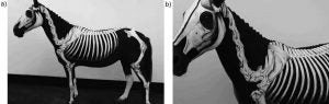
Sattin, Mariana M. et al
2018
Use of a garment as an alternative to body painting in equine musculoskeletal anatomy teaching
Journal of Veterinary Medical Education 45(1):119-125
 |
 |
 |
 |
 |
 |
 |
The fact that garments are amenable to palpation by large groups of students with no damage to the painting favors repeated use in hands-on wet labs. Garments such as the one described in this article introduce a novel approach to interdisciplinary teaching and learning, which can be combined with traditional anatomy teaching methods. … Steps in garment construction are highlighted and indications, advantages, and limitations of the method discussed.
Schoenfeld-Tacher, Regina M. et al
2017
Evaluation of 3D additively manufactured canine brain models for teaching veterinary neuroanatomy
Journal of Veterinary Medical Education 44(4):612-619
 |
 |
 |
Students’ performance on a practical neuroanatomy exam was compared, and no significant differences were found in scores based on the type of material (3D AM models or plastinated specimens) used for instruction. Students in both groups were equally able to identify neuroanatomical structures on cadaveric material, as well as respond to questions involving application of neuroanatomy knowledge. Therefore, we postulate that 3D AM canine brain models are an acceptable alternative to plastinated specimens in teaching veterinary neuroanatomy.
Snell, J.R. et al
1991-01-01
A veterinary digital anatomical database
Proceedings / The … Annual Symposium on Computer Application [sic] in Medical Care 762-765
This paper describes the Veterinary Digital Anatomical Database Project. The purpose of the project is to investigate the construction and use of digitally stored anatomical models. … By using a digital database, the student will have the ability to view and manipulate anatomical structures in ways that are not available through interactive video disk (IVD). IVD constrains the student to preselected views and sections stored on the disk.
Stead, Rachel et al
2021-10-01
Using Play-Doh to Enhance the Perceived Learning of Veterinary Medicine
Journal of Veterinary Medical Education 48(5):549-553
 |
…despite some initial hesitation surrounding the use of what some might perceive as a toy in the higher education classroom, the learners believed that the approach allowed improvement in terms of their understanding, knowledge retention and recall. They reported that the approach enabled greater visualization of the structures they were representing. For teachers, the approach has the advantage that the material is cheap, readily available, easily manipulated, can be reused, and needs no sophisticated technology.
Suñol, Anna et al
2019
Use of three-dimensional printing models for veterinary medical education: Impact on learning how to identify canine vertebral fractures
Journal of Veterinary Medical Education 46(4):523-532
 |
 |
 |
Tamayo-Arango, Lynda J. et al
2020-07-01
Body Painting of the Horse and Cow to Learn Surface Anatomy
Journal of Veterinary Medical Education 47(4):395-401
 |
 |
 |
 |
 |
The results showed that body painting sessions improved learning of anatomical concepts and could serve as a bridge between cadaver anatomy and living animal anatomy.
Tham, Heng L. et al
2023-06-01
A Novel Canine Otoscopy Teaching Model for Veterinary Students
Journal of Veterinary Medical Education 50(3):266-275
 |
 |
 |
Theoret, Christine Laura et al
2007
Why dissection videos should not replace cadaver prosections in the gross veterinary anatomy curriculum: Results from a comparative study
Journal of Veterinary Medical Education 34(2):151-156
In conclusion, while our data suggest that cadaver prosections are superior to video demonstrations, it is apparent that students can learn bovine abdominal anatomy by both methods.
Tiplady, Catherine et al
2011
Veterinary science student preferences for the source of dog cadavers used in anatomy teaching
Alternatives to Laboratory Animals 39(5):461-469
The authors demonstrate that students, although initially having ethical concerns about the source of cadavers, appear to lose this sensitivity as their exposure to consumptive use of animals increases.
These findings are consistent with the hypothesis that veterinary students become more accepting of the euthanasia of unwanted healthy animals for education as they progress through the veterinary programme, in contexts such as the current study. This could occur due to increased acceptance of the euthanasia of healthy animals generally, a decline in moral development, desensitisation, and/or the belief that healthy animal cadavers offer a superior learning experience.
Velásquez, José et al
2023-06-01
Development of an Online Distance Learning Platform Combining Anatomy, Imaging, and Surgical Practice to Support Mastery Learning of the Equine Locomotor Apparatus
Journal of Veterinary Medical Education 50(3):252-257
Yamada, Kazutaka et al
2007
The use of multi–detector row computed tomography (MDCT) as an alternative to specimen preparation for anatomical instruction
Journal of Veterinary Medical Education 34(2):143-150
 |
 |
 |
 |
 |
 |
 |
 |
With this educational support system, anatomical figures can be explained using living animals instead of specimens. In addition, clinical representative examples can be used to show anatomical disorders to students. … This system can enhance veterinary students’ understanding and interest in anatomy and can enable us to offer them a quality veterinary medical education. We concluded that CAD is a useful new option not only for clinical service but also for veterinary education.
Updated 2026-07-16