
It is often argued that anatomy can only be effectively taught through individual student dissection. Such an approach requires an enormous number of cadavers, which puts a strain on any source, even those that are morally defensible. There is no evidence, however, that this approach is educationally necessary, as shown in published studies (some below). If you are aware of other examples you believe to be important to include here, please send the information to HEVM for consideration.
New addition:
HSVMA Educational Memorial Programs
Information on developing a willed-body (ethically sourced cadaver) program
 |
This is an interactive program of two-dimensional images of the dissection of a dog. It was produced by Dr Ray Whalen at the Colorado State University, College of Veterinary Medicine and Biomedical Sciences. Although it is meant to be used in conjunction with traditional dissection, it could be used to enhance a student’s learning through prosections. Part of it is evaluated in Linton et al 2005.
Veterinary Anatomy at the College of Veterinary Medicine, University of Minnesota
This is an online program illustrating various aspects of anatomy.
Multi–detector row computed tomography (MDCT)
 |
 |
 |
Reported by Yamada et al 2007.
The Glass Horse – The Equine Colic CD
 |
Provides normal equine anatomy as well as diseases. From Web site: A comprehensive exploration of the equine abdominal anatomy, the veterinarian’s approach to diagnosis, and detailed 3-D animations depicting more than 25 different diseases.
Reported by Sattin et al 2018.
This is a 3D virtual canine cadaver, part of the Anatomage Table Vet system.
The following includes literature cited above or which is relevant to the issue of anatomy instruction. Some are older citations, demonstrating the usefulness of prosections and related issues. Some concern human medical school education, but the principle findings may have applicability to veterinary medical education. Others may not have practical application, but may be useful in developing arguments to promote humane alternatives by showing student preferences, student sensitivities and the world-wide nature of the concern for humane alternatives. The titles are linked either to a publicly available copy of the document or to a digital object identifier. If there are illustrations which may be publicly viewable, these are also linked, but there is no guarantee that they would be viewable across all platforms.
Adams, D.R. 1974 “The teaching of comparative gross anatomy through the prosection approach” Journal of Veterinary Medical Education 1(1):16-17
As there was insufficient time to dissect much more than the thoracic and pelvic limbs, it was decided to use prosections combined with freshly killed material and live animals, totally discontinuing the dissection of embalmed material.
Al-Khalili, Sereen M. and Coppoc, Gordon L. 2014 2D and 3D stereoscopic videos used as pre-anatomy lab tools improve students’ examination performance in a veterinary gross anatomy course Journal of Veterinary Medical Education 41(1):68-76
Alexander, Justin. 1970 Dissection versus prosection in the teaching of anatomy Journal of Medical Education 45(8):600-606
The statistical results of this study indicate that there were no significant differences in students’ learning which could be attributed to the method of instruction, but that time could be saved if prosection, rather than dissection were to be adopted as the required laboratory procedure.
Despite students’ contention that they can learn more easily [via dissection], this was not borne out.
Allavena, Rachel E.; Schaffer-White, Andrea B.; Long, Hanna and Alawneh, John I. 2017 Technical skills training for veterinary students: A comparison of simulators and video for teaching standardized cardiac dissection Journal of Veterinary Medical Education 44(4):620-631
 |
The major conclusion of this study was that both methods are effective tools for technical skills training, but consideration should be given to the preferred learning style of adult learners to maximize educational outcomes.
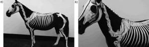
Bietzk, Edward; Weller, Renate; Simons, Victoria and Channon, Sarah B. 2019-09-01 Anatomy Teaching, a “Model” Answer? Evaluating “Geoff”, a Painted Anatomical Horse, as a Tool for Enhancing Topographical Anatomy Learning Anatomical Sciences Education 12(5):529-540
 |
The students found the model to be extremely useful and both groups found the sessions enjoyable. The model will be of benefit as a complementary learning tool for students.
Borgeat, Kieran; Shearn, Andrew I.U.; Payne, Jessie Rose; Hezzell, Melanie and Biglino, Giovanni 2022-06-01 Three-Dimensional Printed Models of the Heart Represent an Opportunity for Inclusive Learning Journal of Veterinary Medical Education 49(3):346-352
 |
Three-dimensional (3D) printed models of anatomic structures offer an alternative to studying manufactured, “idealized” models or cadaveric specimens. … In conclusion, these pilot data suggest that 3D printed canine cardiac models are feasible to produce and represent an inclusive learning opportunity, promoting student engagement.
Bogert, K.; Platt, Simon; Haley, Allison; Kent, Marc; Edwards, Gaylen; Dookwah, H. and Johnsen, Kyle 2016 Development and use of an interactive computerized dog model to evaluate cranial nerve knowledge in veterinary students Journal of Veterinary Medical Education 43(1):26-32
 |
…we have developed a computerized simulated dog head that can exhibit cranial nerve dysfunctions and respond to specific testing procedures in a clinically accurate manner. … In an experiment conducted with 97 freshman veterinary students who had recently been taught cranial nerve anatomy and function, we found that examination performance decreased with the need for interactivity compared to memorization of fact, while satisfaction increased. Students were less likely to identify the correct disorder when they had to conduct the examination of the virtual dog themselves, revealing an inadequacy in traditional neuroanatomical teaching. However, students overwhelmingly supported the use of interactive question for assessment. Interestingly, performance on text-based questions did not correlate significantly with interactive or video questions. The results have implications for veterinary teaching and assessment within the classroom and in clinical environments.
Böttcher, Peter; Maierl, Johann; Schiemann, Thomas; Glaser, Christian; Weller, Renate; Hoehne, Karl-Heinz; Reiser, Maximilian and Liebich, Hans-Georg. 1999 The Visible Animal Project: A three-dimensional, digital database for high quality three-dimensional reconstructions Veterinary Radiology & Ultrasound 40(6):611-616
The “Visible Animal Project” (VAP) is comprised of axial anatomic cryosections and corresponding CT and MR images of a mature dog. The digital database is used for the creation of three-dimensional computer graphics of canine anatomy. The technique of cryodissection is described in detail.
Canright, Abigail; Bescoby, Sam and Dickson, Julie 2023-04-01 Evaluation of a 3D Computer Model of the Equine Paranasal Sinuses as a Tool for Veterinary Anatomy Education Journal of Veterinary Medical Education 50(2):234-242
 |
Findings suggest the 3D-ESM is an effective educational tool that aids in confidence, enjoyment, and knowledge acquisition. Though it was not better than traditional methods in terms of anatomy knowledge exam scores, the model is a valuable inclusion into the veterinary anatomy curriculum.
da Silveira, Erick Eduardo; da Silva Lisboa Neto, Antônio Francisco; Pereira, Helton Carlos Sabino; Ferreira, Janaina Santos; dos Santos, Amilton Cesar; Siviero, Fábio; da Fonseca, Ricardo and de Assis Neto, Antonio Chaves 2021-12-01 Canine Skull Digitalization and Three-Dimensional Printing as an Educational Tool for Anatomical Study Journal of Veterinary Medical Education 48(6):649-655
 |
 |
The results showed that the anatomical structures of the 3D-printed skulls were similar to the real skulls. There was no significant difference between the test scores of the students that did their test using the real skulls and those using 3D prints. In conclusion, it was possible to construct a dynamic and printed digital 3D collection for studies of the comparative anatomy of canine skull species from real skulls, suggesting that 3D-digitalized and-printed skulls can be used as tools in veterinary anatomy teaching.
Daviau, Judith S.; Parker, Jacquelyn C.; Parmelee, Robert H.; Jahn, Sharon E. and Frank, Deborah A. 1997 The use of plastinated specimens as teaching aids of orolaryngeal anatomy in selected laboratory animals Contemporary Topics in Laboratory Animal Science 36(6):50-52
The process of plastination produces stable, durable, maintenance- free specimens that are superior to any other alternative available and also allow for the reduction in the numbers of animals used for teaching.
Davy, Rachel B.; Hamel, Philip E.; Su, Yu; Berry, Clifford R. and Conner, Bobbi J. 2019 Evaluation of two training methods for teaching the Abdominal Focused Assessment with Sonography for Trauma Technique (A-FAST) to first- and second-year veterinary students Journal of Veterinary Medical Education 46(2):258-263
This study compared the results of two training techniques, live-animal training and online video instruction…All participants were assessed on their ability to find and correctly name the four A-FAST quadrants on a live animal. We found a significant difference between the two groups in the students’ ability to identify the diaphragmatic-hepatic (DH) view, but for the other three views (hepatorenal, splenorenal, and cystocolic), training method did not affect performance. Results suggest the potential for using a multi-modal instructional approach to teach ultrasound techniques to veterinary students.
Dickson, Julie; Gardiner, Andrew and Rhind, Susan 2022-06-01 Veterinary Anatomy Education and Spatial Ability: Where Now and Where Next? Journal of Veterinary Medical Education 49(3):297-305
This article situates current understandings of spatial ability in the context of veterinary anatomy education. As in medical education, veterinary courses are increasingly using physical and computer-based models and computer programs to supplement or even replace cadavers. In this article, we highlight the importance of spatial ability in the learning of anatomy and make methodological recommendations for future studies to ensure a robust evidence base is developed.
Elnady, Fawzy A. 2019 Innovative, simple models for teaching neuroanatomy using the Elnady Technique Journal of Veterinary Medical Education 46(2):214-217
 |
 |
 |
 |
 |
…the Elnady Technique…results in realistic, durable, odorless, soft, flexible slices…invaluable for interpretation of clinical imaging modalities, such as computed tomography (CT) and magnetic resonance imaging (MRI). These ethically sourced models can provide a replacement for the killing of animals for practical classes.
Gummery, Erica; Cobb, Kate A.; Mossop, Liz H. and Cobb, Malcolm A. 2018 Student perceptions of veterinary anatomy practical classes: A longitudinal study Journal of Veterinary Medical Education 45(2):163-176
Hoffmann, Darren S.; May, Nikolas; Thomsen, Timothy; Holec, Megan; Andersen, Kathleen H. and Pizzimenti, Marc A. 2010 Medical students using plastinated prosections as a sole learning tool perform equally well on identification exams as compared to those performing dissections over the same regions The FASEB Journal 24(1):176.5
Although involved human medical school students, the principles would apply to veterinary medical school.
These data support use of PP [plastinated prosections] in the medical-level anatomy curriculum, but student response data indicate limitations.
Jones, Norbert A.; Olafson, Raymond P. and Sutin, Jerome. 1978 Evaluation of a gross anatomy program without dissection Journal of Medical Education 53(3):198-205
Students in human medical school studied gross anatomy using a variety of media, including prosections.
…as measured by conventional examinations, students in the multimedia program with prosection tutorials learned human anatomy as well as those in the traditional lecture-dissection program.
Kinnamon, K.K.; Holborow, G.S.; Simmonds, R.C. and Sheridan, M.N. 1992 “A method of preparing veterinary gross anatomy specimens: Multi-year holdings and use requires no refrigeration” Journal of Veterinary Medical Education 19(4):108-110
Kovarik, M. Cathleen and Hancock, Tamara S. 2023-08-01 Effectiveness of a Student-Developed Instructional Video in Learning the Anatomy of the Equine Distal Limb Journal of Veterinary Medical Education 50(4):457-462
 |
Kozlowski, G.P. and St. Clair, L.E. 1974 “Use of freeze-dried specimens in the teaching of veterinary gross anatomy” Journal of Veterinary Medical Education 1(1):1-3
The preparation and use of freeze-dried specimens in teaching gross veterinary anatomy are described. Examples are given of how easily handled, cross-sectioned specimens can help students visualize and identify anatomic structures.
Küçükaslan, Özgül; Erdogan, Serkan and Bulut, Ilhami 2019 Turkish undergraduate veterinary students’ attitudes to use of animals and other teaching alternatives for learning anatomy Journal of Veterinary Medical Education 46(1):116-127
Kumar, Amarendhra; Murtaugh, Robert; Brown, Donald; Ballas, True; Clancy, Elizabeth and Patronek, Gary 2001 Client donation program for acquiring dogs and cats to teach veterinary gross anatomy Journal of Veterinary Medical Education 28(2):73-77
Describes the donor-based program for acquiring cadavers at Tufts University.
Latorre, Rafael M.; García-Sanz, Mari P.; Moreno, Matilde; Hernández, Fuensanta; Gil, Francisco; López, Octavio; Ayala, Maria D.; Ramírez, Gregorio; Vázquez, Jose M.; Arencibia, Alberto and Henry, Robert W. 2007 How useful is plastination in learning anatomy? Journal of Veterinary Medical Education 34(2):172-176
Latorre, Rafael; Bainbridge, David; Tavernor, Angie and Albors, Octavio López 2016 Plastination in anatomy learning: An experience at Cambridge University Journal of Veterinary Medical Education 43(3):226-234
Describes their experience using plastination alongside traditional ‘wet’ dissections.
Leandro, Rafael M.; Filho, Roberto P.P. Foz; De Silvio, Mauricio M.; Quilici, Ana P.; Sattin, Mariana M.; Paretsis, Barbara F. and Souza, Vanessa A. 2019 Construction of the equine digestive system: A tool for teaching topographical anatomy Journal of Veterinary Medical Education 46(1):108-115
 |
 |
 |
 |
 |
The method described in this study is a low-cost, user-friendly teaching tool that is potentially applicable across different academic disciplines and that can also be used to construct models of other systems and species. However, it should be combined with other anatomy teaching methods because it does not provide detailed representation of specific digestive system organ features.
Linton, Andrea; Schoenfeld-Tacher, Regina and Whalen, L. Ray 2005 Developing and implementing an assessment method to evaluate a Virtual Canine Anatomy program Journal of Veterinary Medical Education 32(2):249-254
Martinsen, Siri and Jukes, Nick 2007 Ethically sourced animal cadavers and tissue: Considerations for education and training Alternatives to Animal Testing and Experimentation 14:265-268
Discusses the ethical and practical aspects of using cadavers, particularly in veterinary medical anatomy and surgical training.
Nemanic, Sarah; Mills, Serena; Viehdorfer, Matt; Clark, Terri and Bailey, Mike 2016 The effectiveness of a 3D computerized tutorial to enhance learning of the canine larynx and hyoid apparatus Journal of Veterinary Medical Education 43(3):243-254
 |
 |
 |
 |
 |
 |
This study demonstrates that a 3D computerized tutorial is more effective in teaching the anatomy of the canine hyoid apparatus and larynx than 3D images without a tutorial. Students likewise rated their learning experience higher when using the 3D computerized tutorial.
Preece, Daniel; Williams, Sarah B.; Lam, Richard and Weller, Renate 2013 “Let’s get physical”: Advantages of a physical model over 3D computer models and textbooks in learning imaging anatomy Anatomical Sciences Education 6(4):216-224
 |
Three-dimensional (3D) information plays an important part in medical and veterinary education. … Our results suggest that physical models may hold a significant advantage over alternative learning resources in enhancing visuospatial and 3D understanding of complex anatomical architecture, and that 3D computer models have significant limitations with regards to 3D learning.
Prentice, Ernest D.; Metcalf, William K.; Quinn, Thomas H.; Sharp, John G.; Jensen, Richard H. and Holyoke, Edward A. 1977 Stereoscopic anatomy: evaluation of a new teaching system in human gross anatomy Journal of Medical Education 52(9):758-763
Evaluation data suggest that this program, while having minor limitations in terms of anatomical orientation, does provide a viable alternative to dissection.
Provo, Judy A. and Lamar, Carlton, H. 1995 Prosection as an approach to student-centered learning in veterinary gross anatomy Journal of the American Veterinary Medical Association 206(2):158-161
By using prosections (as opposed to having each student or pair of students dissect their own cadaver) and increasing the number of students studying a particular cadaver, the overall number of animals used is decreased. No difference in learning is seen or demonstrable.
Analysis of performance in this course [in which prosection was compared with dissection] showed that veterinary students do not need to dissect every area to learn comparative anatomy effectively. Students made as many errors on structures that they learned by dissection as they did on structures learned from peer prosection. This finding is important for 2 reasons. First, learning by demonstration requires much less class time than does dissection (2 hours for a dissection assignment vs 20 to 30 minutes for a demonstration). Second, the use of prosections can reduce the number of animals needed to teach gross anatomy.
These findings are in agreement with those in human anatomy. …studies measuring retention have shown no difference between prosection and dissection.
Sattin, Mariana M.; Silva, Vickitoriana K.A.; Leandro, Rafael M.; Filho, Roberto P.P. Foz and De Silvio, Mauricio M. 2018 Use of a garment as an alternative to body painting in equine musculoskeletal anatomy teaching Journal of Veterinary Medical Education 45(1):119-125
 |
 |
 |
 |
 |
 |
 |
The fact that garments are amenable to palpation by large groups of students with no damage to the painting favors repeated use in hands-on wet labs. Garments such as the one described in this article introduce a novel approach to interdisciplinary teaching and learning, which can be combined with traditional anatomy teaching methods. … Steps in garment construction are highlighted and indications, advantages, and limitations of the method discussed.
Schoenfeld-Tacher, Regina M.; Horn, Timothy J.; Scheviak, Tyler A.; Royal, Kenneth D. and Hudson, Lola C. 2017 Evaluation of 3D additively manufactured canine brain models for teaching veterinary neuroanatomy Journal of Veterinary Medical Education 44(4):612-619
 |
 |
 |
Students’ performance on a practical neuroanatomy exam was compared, and no significant differences were found in scores based on the type of material (3D AM models or plastinated specimens) used for instruction. Students in both groups were equally able to identify neuroanatomical structures on cadaveric material, as well as respond to questions involving application of neuroanatomy knowledge. Therefore, we postulate that 3D AM canine brain models are an acceptable alternative to plastinated specimens in teaching veterinary neuroanatomy.
Stead, Rachel; Lygo-Baker, Simon; Coppi, Antonio Augusto and De Melo, Mariana Pereira 2021-10-01 Using Play-Doh to Enhance the Perceived Learning of Veterinary Medicine Journal of Veterinary Medical Education 48(5):549-553
 |
…despite some initial hesitation surrounding the use of what some might perceive as a toy in the higher education classroom, the learners believed that the approach allowed improvement in terms of their understanding, knowledge retention and recall. They reported that the approach enabled greater visualization of the structures they were representing. For teachers, the approach has the advantage that the material is cheap, readily available, easily manipulated, can be reused, and needs no sophisticated technology.
Suñol, Anna; Aige, Vicente; Morales, Carles; López-Beltran, Marta; Feliu-Pascual, Alejandro Luján and Puig, Jordi 2019 Use of three-dimensional printing models for veterinary medical education: Impact on learning how to identify canine vertebral fractures Journal of Veterinary Medical Education 46(4):523-532
 |
 |
 |
Tamayo-Arango, Lynda J. and Mejía-Durango, María A. 2020-07-01 Body Painting of the Horse and Cow to Learn Surface Anatomy Journal of Veterinary Medical Education 47(4):395-401
 |
 |
 |
 |
 |
The results showed that body painting sessions improved learning of anatomical concepts and could serve as a bridge between cadaver anatomy and living animal anatomy.
Tham, Heng L.; Elnady, Fawzy A. and Byrnes, Meghan K. 2023-06-01 A Novel Canine Otoscopy Teaching Model for Veterinary Students Journal of Veterinary Medical Education 50(3):266-275
 |
 |
 |
Theoret, Christine Laura; Carmel, Éric-Norman and Bernier, Sonia 2007 Why dissection videos should not replace cadaver prosections in the gross veterinary anatomy curriculum: Results from a comparative study Journal of Veterinary Medical Education 34(2):151-156
In conclusion, while our data suggest that cadaver prosections are superior to video demonstrations, it is apparent that students can learn bovine abdominal anatomy by both methods.
Tiplady, Catherine; Lloyd, Shan and Morton, John 2011 Veterinary science student preferences for the source of dog cadavers used in anatomy teaching Alternatives to Laboratory Animals 39(5):461-469
The authors demonstrate that students, although initially having ethical concerns about the source of cadavers, appear to lose this sensitivity as their exposure to consumptive use of animals increases.
These findings are consistent with the hypothesis that veterinary students become more accepting of the euthanasia of unwanted healthy animals for education as they progress through the veterinary programme, in contexts such as the current study. This could occur due to increased acceptance of the euthanasia of healthy animals generally, a decline in moral development, desensitisation, and/or the belief that healthy animal cadavers offer a superior learning experience.
Velásquez, José; da Silva, Luis Lopes Correia and Miglino, Maria Angélica 2023-06-01 Development of an Online Distance Learning Platform Combining Anatomy, Imaging, and Surgical Practice to Support Mastery Learning of the Equine Locomotor Apparatus Journal of Veterinary Medical Education 50(3):252-257
Yamada, Kazutaka; Taniura, Tokunori; Tanabe, Shigeyuki; Yamaguchi, Miho; Azemoto, Shogo and Wisner, Erik R. 2007 The use of multi–detector row computed tomography (MDCT) as an alternative to specimen preparation for anatomical instruction Journal of Veterinary Medical Education 34(2):143-150
 |
 |
 |
 |
 |
 |
 |
 |
With this educational support system, anatomical figures can be explained using living animals instead of specimens. In addition, clinical representative examples can be used to show anatomical disorders to students. … This system can enhance veterinary students’ understanding and interest in anatomy and can enable us to offer them a quality veterinary medical education. We concluded that CAD is a useful new option not only for clinical service but also for veterinary education.
Updated 2023-12-14